Case 102
Jump to navigation
Jump to search
| Courtesy of: AMC Echolab, AMC, The Netherlands | |

|

|
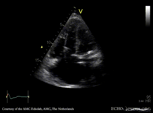
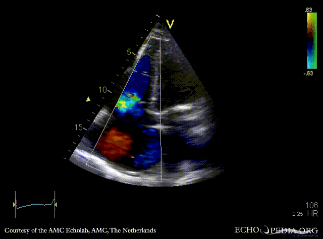
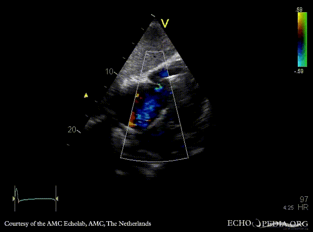
| A4CH: dilated right ventricle and right atrium, pacemaker lead in situ | A4CH with Color Doppler: severe tricuspid regurgitation |

|

|
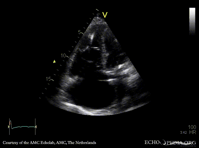
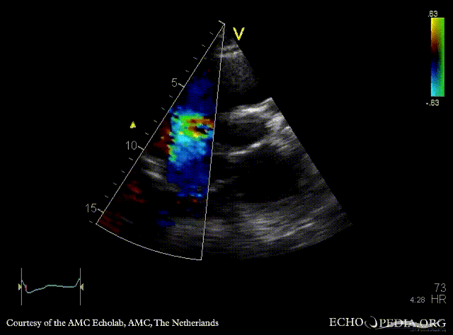
| A4CH: coronary sinus a vue | PSAX with Color Doppler: severe tricuspid regurgitation |
 |

|
| Continuous-wave signal of tricuspid regurgitation | Subcostal view: severe tricuspid regurgitation |
 |
 |
| Dilated vena cava inferior, no diameter variations during respiration | Pulsed-wave Doppler signal of hepatic veins: systolic flow reversal |