Tricuspid
Anatomy
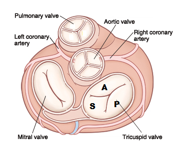
The tricuspid valve, ensures that there is no backflow of blood from the right ventricle to the right atrium. The tricuspid valve is like the Latin name indicates consists of three leaflets: the septal (S), the anterior (A) and posterior (P) leaflet, whose anterior leaflet is the largest. The tricuspid valve, distinguishes itself not only by the number of valve leaflets of the mitral valve, but also by the manner of attachment. The chordae of the mitral valve attached to two papillary cups which the tricuspid valve attached to a lot more muscular and cups directly to the septum. The valve is located slightly to the apex than the mitral valve.[1]
Echocardiographic views
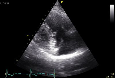
|
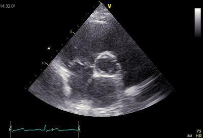
|
| Plax by tilted | PSax Ao |
|---|---|
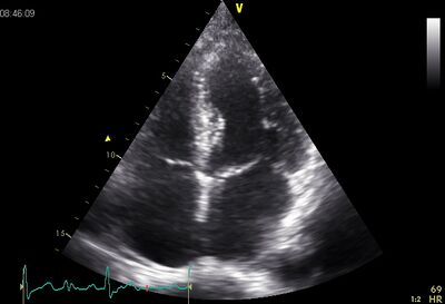
|
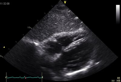
|
| AP4CH | Subcostal |
Stenosis
An isolated tricuspidaalklepstenose is extremely rare in adulthood. Almost always there is a combination with a more or less serious tricuspidaalklepinsufficiëntie. When a mitral valve stenosis in old age the cause is almost always a previously created rheumatic fever. In 10% of the patients, also, there appeared to be a tricuspidaalklepstenose. Dooming the valve leaflets is always an expression of a tricuspidaalklepstenose. For assessing the severity of a tricuspidaalklepstenose are no objective criteria exist, however it is important to realize that the pressure differences tricuspidaalklepstenose be less. At a mean gradient of 5 or greater may also be a serious tricuspidaalklepstenose be. The peak gradient is high, both in patients with a severe stenosis without TI as in patients with severe TS with low TI. The deceleration time will be obvious differences.
Click here for quantification tricuspid valve stenosis.
