Case 138
Jump to navigation
Jump to search
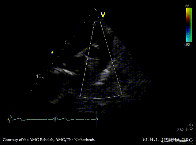
| Courtesy of: AMC Echolab, AMC, The Netherlands | |
| <html5media height="350" width="279" autoplay="true" loop="true">File:E00727.mp4</html5media> | <html5media height="350" width="279" autoplay="true" loop="true">File:E00728.mp4</html5media> |
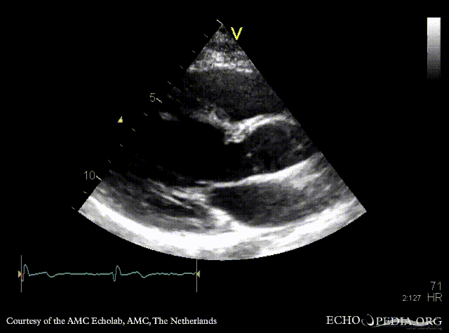
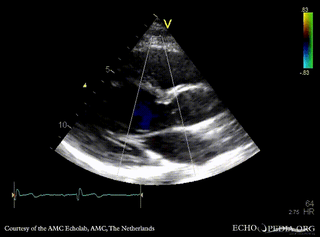
| PSAX: subvalvular membrane | PSAX with Color Doppler: high velocity flow in LVOT |
| <html5media height="350" width="279" autoplay="true" loop="true">File:E00729.mp4</html5media> | <html5media height="350" width="279" autoplay="true" loop="true">File:E00730.mp4</html5media> |
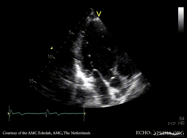
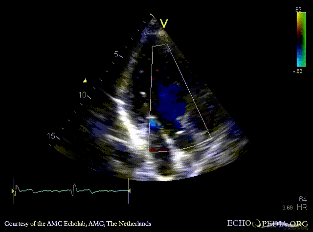
| A3CH: subvalvular membrane | A3CH with Color Doppler: high velocity flow in LVOT |
 |
 |
| Continuous-wave Doppler signal of transaortic flow | Pulsed-wave Doppler signal of LVOT flow: mild dynamic gradient |
| <html5media height="350" width="279" autoplay="true" loop="true">File:E00733.mp4</html5media> |  |
| Suprasternal view: high velocity flow in ascending aorta | Continuous-wave Doppler signal of flow in ascending aorta |