Case 66 and Case 20: Difference between pages
(Difference between pages)
Jump to navigation
Jump to search
Secretariat (talk | contribs) (Created page with '{{EchoCase |Title = Severe pulmonary regurgitation |CasePresentation = |Courtesy = AMC Echolab, AMC, The Netherlands |filepointer1=<flash>file=E00380.swf|quality=best|align=...') |
Secretariat (talk | contribs) No edit summary |
||
| Line 1: | Line 1: | ||
{{EchoCase | {{EchoCase | ||
|Title = | |Title = Amyloidosis | ||
|CasePresentation = | |CasePresentation = | ||
|Courtesy = [[AMC Echolab, AMC, The Netherlands]] | |Courtesy = [[AMC Echolab, AMC, The Netherlands]] | ||
|filepointer1=<flash>file= | |filepointer1=<flash>file=E00192.swf|quality=best|align=center|width=350|height=279</flash> | ||
|file_name1= | |file_name1=E00192 | ||
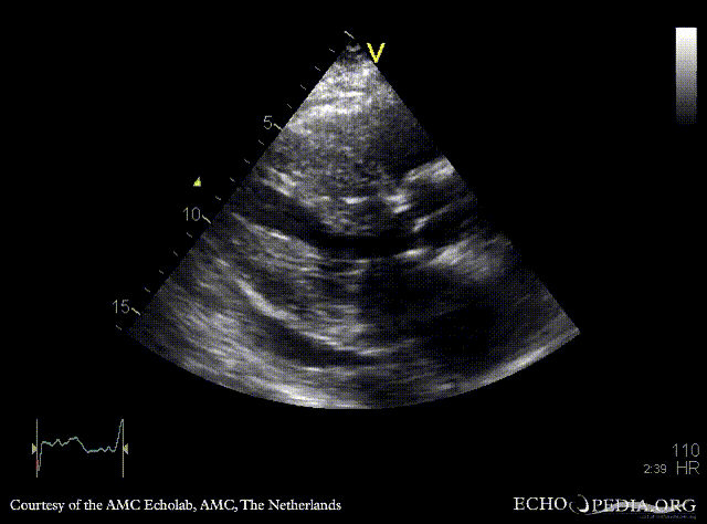
|descriptionfile1= | |descriptionfile1=PLAX: concentric left ventricle hypertrophy and pericardial effusion in patient with amyloidosis | ||
|filepointer2= | |filepointer2=<flash>file=E00193.swf|quality=best|align=center|width=350|height=279</flash> | ||
|file_name2= | |file_name2=E00193 | ||
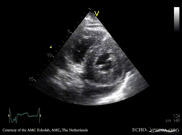
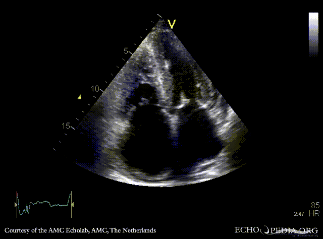
|descriptionfile2= | |descriptionfile2=PSAX: concentric left and right ventricle hypertrophy | ||
|filepointer3=<flash>file= | |filepointer3=<flash>file=E00194.swf|quality=best|align=center|width=350|height=279</flash> | ||
|file_name3= | |file_name3=E00194 | ||
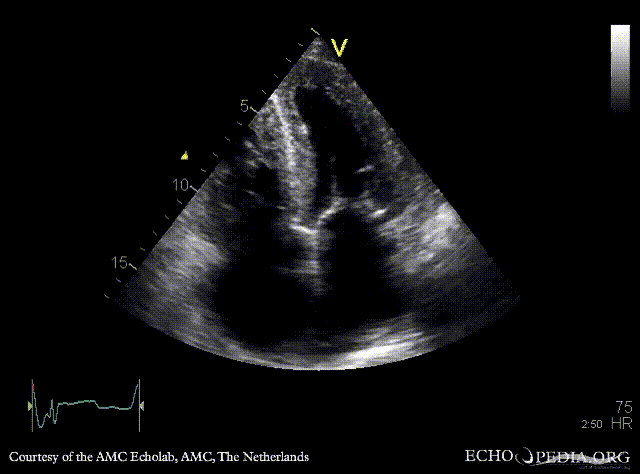
|descriptionfile3=A4CH: | |descriptionfile3=A4CH: concentric left and right ventricle hypertrophy, biatrial enlargement | ||
|filepointer4=<flash>file=E00195.swf|quality=best|align=center|width=350|height=279</flash> | |||
|file_name4=E00195 | |||
|descriptionfile4=A4CH: moderate mitral regurgitation | |||
|filepointer5=<flash>file=E00196.swf|quality=best|align=center|width=350|height=279</flash> | |||
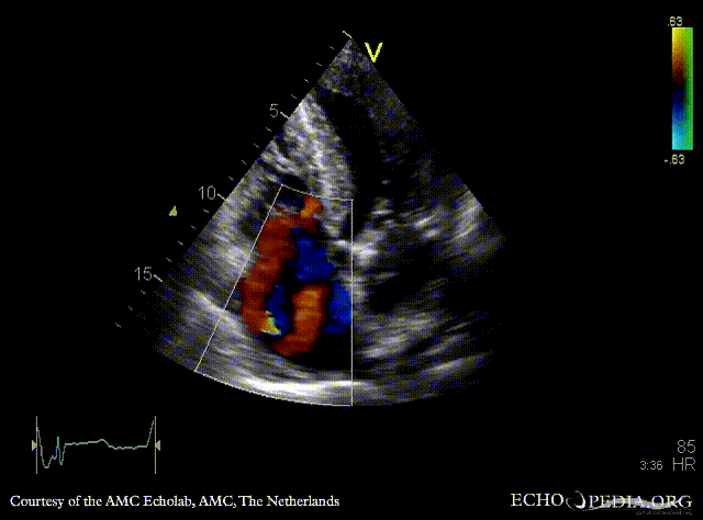
|file_name5=E00196 | |||
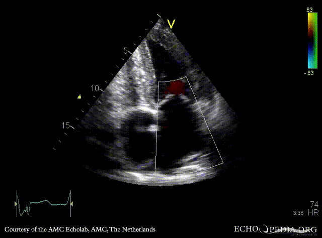
|descriptionfile5=A4CH: severe tricuspid regurgitation | |||
|filepointer6=[[File:E00197.jpg|350px|left]] | |||
|file_name6= | |||
|descriptionfile6=Pulsed-wave Doppler signal: restrictive pattern of mitral inflow | |||
|filepointer7=<flash>file=E00198.swf|quality=best|align=center|width=350|height=279</flash> | |||
|file_name7=E00198 | |||
|descriptionfile7=A4CH: concentric left and right ventricle hypertrophy, biatrial enlargement and diffuse mitral valve thickening | |||
}} | }} | ||
Latest revision as of 18:13, 26 November 2009
| Courtesy of: AMC Echolab, AMC, The Netherlands | |
| <flash>file=E00192.swf | <flash>file=E00193.swf |
| PLAX: concentric left ventricle hypertrophy and pericardial effusion in patient with amyloidosis | PSAX: concentric left and right ventricle hypertrophy |
| <flash>file=E00194.swf | <flash>file=E00195.swf |
| A4CH: concentric left and right ventricle hypertrophy, biatrial enlargement | A4CH: moderate mitral regurgitation |
| <flash>file=E00196.swf |  |
| A4CH: severe tricuspid regurgitation | Pulsed-wave Doppler signal: restrictive pattern of mitral inflow |
| <flash>file=E00198.swf | |
| A4CH: concentric left and right ventricle hypertrophy, biatrial enlargement and diffuse mitral valve thickening | |