Case 23 and Case 22: Difference between pages
(Difference between pages)
Jump to navigation
Jump to search
No edit summary |
Secretariat (talk | contribs) (Created page with '{{EchoCase |Title = Amyloidosis |CasePresentation = |Courtesy = AMC Echolab, AMC, The Netherlands |filepointer1=<flash>file=E00204.swf|quality=best|align=center|width=300|he...') |
||
| Line 1: | Line 1: | ||
{{EchoCase | {{EchoCase | ||
|Title = | |Title = Amyloidosis | ||
|CasePresentation = | |CasePresentation = | ||
|Courtesy = [[AMC Echolab, AMC, The Netherlands]] | |Courtesy = [[AMC Echolab, AMC, The Netherlands]] | ||
|filepointer1=< | |filepointer1=<flash>file=E00204.swf|quality=best|align=center|width=300|height=300</flash> | ||
|file_name1= | |file_name1=E00204 | ||
|descriptionfile1= | |descriptionfile1=PLAX: concentric left ventricle hypertrophy and pericardial effusion in a patient with amyloidosis | ||
|filepointer2=< | |filepointer2=<flash>file=E00205.swf|quality=best|align=center|width=300|height=300</flash> | ||
| | |file_name2=E00205 | ||
| | |descriptionfile2=PSAX: concentric left and right ventricle hypertrophy and pericardial effusion | ||
|filepointer3=<flash>file=E00206.swf|quality=best|align=center|width=300|height=300</flash> | |||
|file_name3=E00206 | |||
|descriptionfile3=PSAX: diffuse thickening of mitral valve, pericardial effusion | |||
|filepointer4=<flash>file=E00207.swf|quality=best|align=center|width=300|height=300</flash> | |||
|file_name4=E00207 | |||
|descriptionfile4=PSAX: diffuse thickening of aortic valve | |||
|filepointer5=<flash>file=E00208.swf|quality=best|align=center|width=300|height=300</flash> | |||
|file_name5=E00208 | |||
|descriptionfile5=PLAX: Color Doppler signal shows mild aortic regurgitation | |||
|filepointer6=[[File:E00209.jpg|300px|left]] | |||
|file_name6= | |||
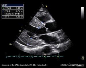
|descriptionfile6=M-Mode through left ventricle | |||
|filepointer7=<flash>file=E00210.swf|quality=best|align=center|width=300|height=300</flash> | |||
|file_name7=E00210 | |||
|descriptionfile7=A4CH: concentric left and right ventricle hypertrophy with reduced systolic function, biatrial enlargement and pericardial effusion | |||
|filepointer8=<flash>file=E00211.swf|quality=best|align=center|width=300|height=300</flash> | |||
|file_name8=E00211 | |||
|descriptionfile8=A2CH: concentric left ventricle hypertrophy | |||
|filepointer9=<flash>file=E00212.swf|quality=best|align=center|width=300|height=300</flash> | |||
|file_name9=E00212 | |||
|descriptionfile9=A3CH: concentric left ventricle hypertrophy | |||
|filepointer10=<flash>file=E00213.swf|quality=best|align=center|width=300|height=300</flash> | |||
|file_name10=E00213 | |||
|descriptionfile10=Subcostal view: concentric left and right ventricle hypertrophy | |||
}} | }} | ||
Revision as of 12:12, 26 November 2009
| Courtesy of: AMC Echolab, AMC, The Netherlands | |
| <flash>file=E00204.swf | <flash>file=E00205.swf |
| PLAX: concentric left ventricle hypertrophy and pericardial effusion in a patient with amyloidosis | PSAX: concentric left and right ventricle hypertrophy and pericardial effusion |
| <flash>file=E00206.swf | <flash>file=E00207.swf |
| PSAX: diffuse thickening of mitral valve, pericardial effusion | PSAX: diffuse thickening of aortic valve |
| <flash>file=E00208.swf | |
| PLAX: Color Doppler signal shows mild aortic regurgitation | M-Mode through left ventricle |
| <flash>file=E00210.swf | <flash>file=E00211.swf |
| A4CH: concentric left and right ventricle hypertrophy with reduced systolic function, biatrial enlargement and pericardial effusion | A2CH: concentric left ventricle hypertrophy |
| <flash>file=E00212.swf | <flash>file=E00213.swf |
| A3CH: concentric left ventricle hypertrophy | Subcostal view: concentric left and right ventricle hypertrophy |
