Tricuspid Insufficiency
Tricuspid insufficiency
Tricuspid insufficiency (TI) is common. If there is a significant mitral valvular insufficiency is present, the sonographer must also look at. To the tricuspid valve TI is also seen in many RV pacemakers, annulus dilatation obv RV dilatation with ASD, pulmonary hypertension, pulmonary embolism and RV infarction. Other causes which there occurs TI's include prolapse, endocarditis, carcinoid, congenital (atrioventricular septal defect, tricuspid atresia). A rare congenital abnormality of the tricuspid valve in which there is present M.Ebstein TI.
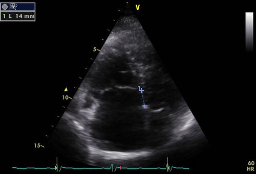
M. Ebstein
In M. Ebstein's tricuspid not well developed. In developing the release the inner layer of the RV wall so that it can form. Tricuspid In M. Ebstein is loslatingsproces not finished making the leaflets still remain glued to the wall. The insertion of the septal and posterior leaflets of the tricuspid valve is M. Ebstein moved more to the apical and anterior leaflet is extra large with many fenestrations. Diagnostic is the distance between the attachment of the septal leaflet to the anterior leaflet of mitral valve ≥ 8mm / m2 (BSA indexed) an indication of M.Ebstein. M. Ebstein may be associated with other congenital abnormalities such as ASD, VSD, tetralogy of Fallot and coarctation of the aorta. In a M. Ebstein's the RV and RA dilated o.b.v. volume load resulting from TI.
Tricuspid regurgitation quantification
| Slight | Moderate | Severe | |
|---|---|---|---|
| RV , RA , VCI dimensions | normal | normal/dilated | dilated |
| Leverveneflow | Systolic dominant | Systolic decrease | Systolic reversal |
| Vena contracta | <7mm | >7mm | |
| PISA | <0.6cm2 | 0.6- 0.9cm2 | >0.9cm2 |
| Density contour + CW signal | light + parabolic | holds + variable | holds + triangular |
| * TI jet / RA area | <20 % low | 20-34% moderately | >35% severe |
| * The size of the color - Doppler surface into right atrium gives an overall impression.[1] | |||
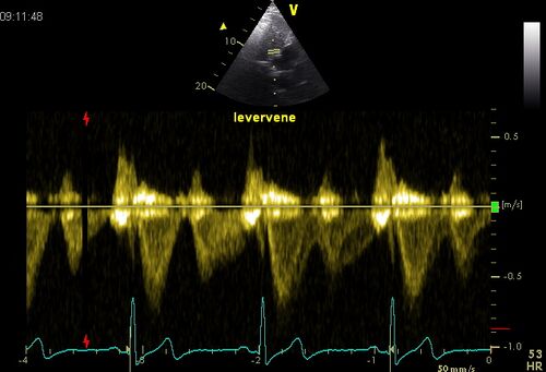
Hepatic vein flow
Pulsed Doppler signal of hepatic vein in severe tricuspid insufficiency.
Holosystolic retrograde flow in the hepatic vein indicates TI hemodynamically important. At greatly increased RA retrograde flow may be lacking in the hepatic vein, despite serious Tl.

|

|
| Normal hepatic vein flow | Hepatic vein systolic flow reversal |
|---|
Vena contracta
The vena contracta TI jet can be used as a measure of the severity of the TI. If the vena contracta diameter more than the 7.5 mm is certainly a serious TI. At a diameter of the vena contracta of less than 6mm is almost certainly no serious TI.
References
- Lancellotti P, Moura L, Pierard LA, Agricola E, Popescu BA, Tribouilloy C, Hagendorff A, Monin JL, Badano L, Zamorano JL, and European Association of Echocardiography. European Association of Echocardiography recommendations for the assessment of valvular regurgitation. Part 2: mitral and tricuspid regurgitation (native valve disease). Eur J Echocardiogr. 2010 May;11(4):307-32. DOI:10.1093/ejechocard/jeq031 |
