Aorta
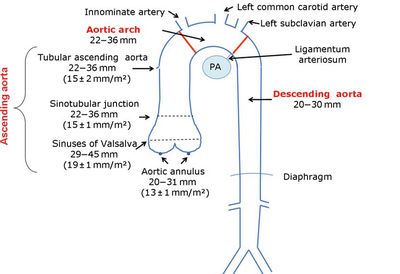
Aortic Dimensions
| Aortic diameters (BSAindex) | |
|---|---|
| Aortic annulus | 20 - 31mm (13 ± 1mm/m2) |
| Aortic Root | 29 - 45mm (19 ± 1mm/m2) |
| Sinotubular junction | 22 - 36mm (15 ± 1mm/m2) |
| Tube | 22 - 36mm (15 ± 2mm/m2) |
| Aortic Arch | 22 -36mm |
| Descending aorta | 20 -30mm |
| Abdominal aorta | 20 -30mm |
| NB. In contrast to tomographic methods, the echocardiographic derived measures are reported as internal diameters. "Recommendations for Aortic Imaging Techniques to Determine the Presence and Progression of Thoracic Aortic Disease".[1] | |

|
| Picture source: European Journal of Echocardiography (2010 ) 11 , 645-658[2] |
|---|
Aortic dissection
Diagnostic is an undulating motion intimal flap, which in more recordings and directions must be seen. The flap should have a movement that is not parallel with any other cardio-thoracic structure.
| Upon dissection watch: |
|
|---|
It also shows the intramural hematoma of the aorta to be aware of the aortic dissection. One variant This does not intraluminal flap was observed making the diagnosis is difficult to establish. Echocardiographic is viewed as a thickened aortic wall.
| Differentiation between true and false lumen: |
|
|---|
Aortic coarctation
Imaging of the aortic arch usually works best from the jugular (sternal supra). When evaluating a patient with a suspected coarctation always pay attention to associated anomalies such as:
- Bicuspid aortic valve
- Aortic valve stenosis
- Patent ductus arteriosus
- VSD
- Mitral valve abnormalities
Determining coarctation[2]
| Instrument | Remark | |
|---|---|---|
| Location | Color doppler | The origin of the carotid and subclavian artery are reference points for locating the coarctation. |
| Speed Profile | Continuous wave | Remember that collaterals systolic maximum speed but does reduce the diastolic gradient persists. In the presence of diastolic forward flow refers to a hemodynamically significant coarctation.
Typical CW Doppler signal from descending aorta with diastolic forward flow matching hemodynamically significant coarctation. |
References
- Hiratzka LF, Bakris GL, Beckman JA, Bersin RM, Carr VF, Casey DE Jr, Eagle KA, Hermann LK, Isselbacher EM, Kazerooni EA, Kouchoukos NT, Lytle BW, Milewicz DM, Reich DL, Sen S, Shinn JA, Svensson LG, Williams DM, American College of Cardiology Foundation/American Heart Association Task Force on Practice Guidelines, American Association for Thoracic Surgery, American College of Radiology, American Stroke Association, Society of Cardiovascular Anesthesiologists, Society for Cardiovascular Angiography and Interventions, Society of Interventional Radiology, Society of Thoracic Surgeons, and Society for Vascular Medicine. 2010 ACCF/AHA/AATS/ACR/ASA/SCA/SCAI/SIR/STS/SVM guidelines for the diagnosis and management of patients with Thoracic Aortic Disease: a report of the American College of Cardiology Foundation/American Heart Association Task Force on Practice Guidelines, American Association for Thoracic Surgery, American College of Radiology, American Stroke Association, Society of Cardiovascular Anesthesiologists, Society for Cardiovascular Angiography and Interventions, Society of Interventional Radiology, Society of Thoracic Surgeons, and Society for Vascular Medicine. Circulation. 2010 Apr 6;121(13):e266-369. DOI:10.1161/CIR.0b013e3181d4739e |
- Evangelista A, Flachskampf FA, Erbel R, Antonini-Canterin F, Vlachopoulos C, Rocchi G, Sicari R, Nihoyannopoulos P, Zamorano J, European Association of Echocardiography, Document Reviewers:, Pepi M, Breithardt OA, and Plonska-Gosciniak E. Echocardiography in aortic diseases: EAE recommendations for clinical practice. Eur J Echocardiogr. 2010 Sep;11(8):645-58. DOI:10.1093/ejechocard/jeq056 |