Case 122: Difference between revisions
Jump to navigation
Jump to search
Secretariat (talk | contribs) (Created page with '{{EchoCase |Title = Pericarditis constrictiva |CasePresentation = |Courtesy = AMC Echolab, AMC, The Netherlands |filepointer1=<flash>file=E00663.swf|quality=best|align=cente...') |
m (Replace html5media with gif) |
||
| (One intermediate revision by one other user not shown) | |||
| Line 4: | Line 4: | ||
|Courtesy = [[AMC Echolab, AMC, The Netherlands]] | |Courtesy = [[AMC Echolab, AMC, The Netherlands]] | ||
|filepointer1= | |filepointer1=[[File:E00663.gif|350px]] | ||
|file_name1=E00663 | |file_name1=E00663 | ||
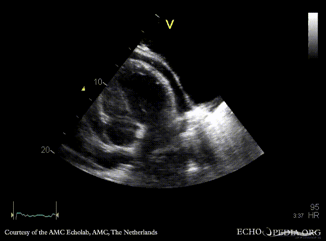
|descriptionfile1=A4CH: pericardial effusion | |descriptionfile1=A4CH: pericardial effusion | ||
| Line 12: | Line 12: | ||
|descriptionfile2=Pulsed-wave Doppler signal of mitral inflow: significant inflow variations during respiration | |descriptionfile2=Pulsed-wave Doppler signal of mitral inflow: significant inflow variations during respiration | ||
|filepointer3= | |filepointer3=[[File:E00665.gif|350px]] | ||
|file_name3=E00665 | |file_name3=E00665 | ||
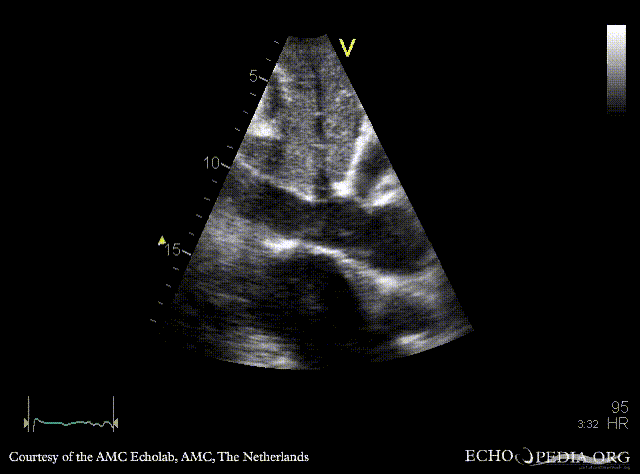
|descriptionfile3=Subcostal view: dilated vena cava inferior, no respiratory variations | |descriptionfile3=Subcostal view: dilated vena cava inferior, no respiratory variations | ||
Latest revision as of 19:52, 1 December 2023
| Courtesy of: AMC Echolab, AMC, The Netherlands | |

|
 |
| A4CH: pericardial effusion | Pulsed-wave Doppler signal of mitral inflow: significant inflow variations during respiration |

|
 |
| Subcostal view: dilated vena cava inferior, no respiratory variations | M-Mode through vena cava inferior |