Case 21: Difference between revisions
Jump to navigation
Jump to search
Secretariat (talk | contribs) No edit summary |
Secretariat (talk | contribs) No edit summary |
||
| Line 6: | Line 6: | ||
|filepointer1=<flash>file=E00199.swf|quality=best|align=center|width=350|height=279</flash> | |filepointer1=<flash>file=E00199.swf|quality=best|align=center|width=350|height=279</flash> | ||
|file_name1=E00199 | |file_name1=E00199 | ||
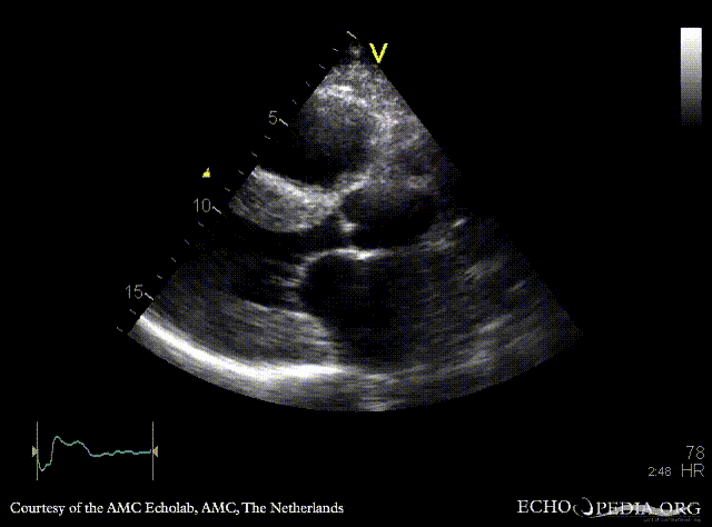
|descriptionfile1= | |descriptionfile1=PLAX: concentric left ventricle hypertrophy in a patient with amyloidosis | ||
|filepointer2=<flash>file=E00200.swf|quality=best|align=center|width=350|height=279</flash> | |filepointer2=<flash>file=E00200.swf|quality=best|align=center|width=350|height=279</flash> | ||
Revision as of 06:34, 11 December 2009
| Courtesy of: AMC Echolab, AMC, The Netherlands | |
| <flash>file=E00199.swf | <flash>file=E00200.swf |
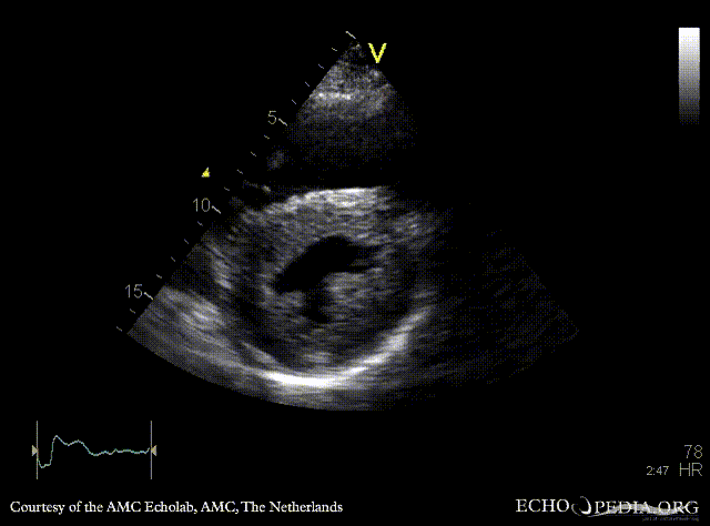
| PLAX: concentric left ventricle hypertrophy in a patient with amyloidosis | PSAX: concentric left and right ventricle hypertrophy |
| <flash>file=E00201.swf | <flash>file=E00202.swf |
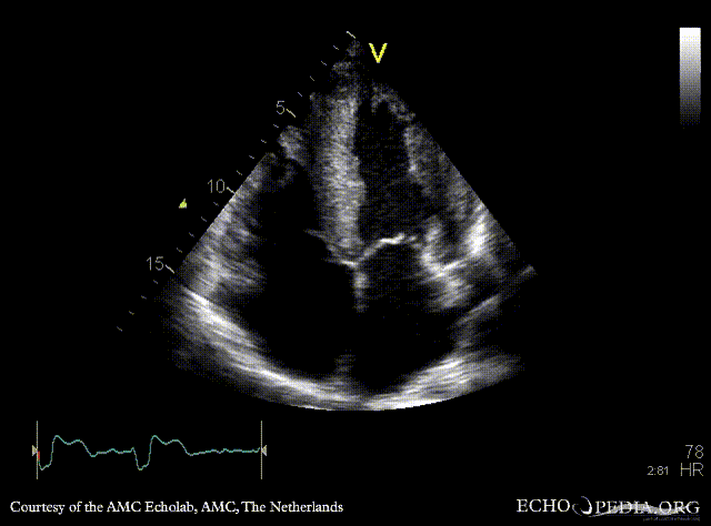
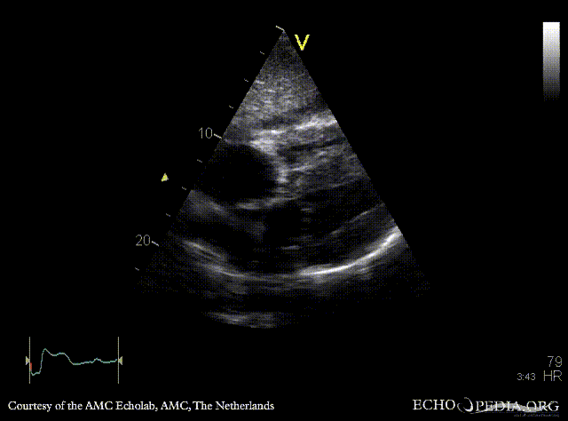
| A4CH: concentric left and right ventricle hypertrophy with reduced systolic function, biatrial enlargement | Subcostal view: concentric left and right ventricle hypertrophy |
 |
|
| Pulsed-wave Doppler signal: restrictive pattern of mitral inflow | |