Cases and Examples: Difference between revisions
Jump to navigation
Jump to search
No edit summary |
No edit summary |
||
| Line 10: | Line 10: | ||
The apical 4 chamber echocardiogram shows severe pericardiual effusion and a swinging heart. | The apical 4 chamber echocardiogram shows severe pericardiual effusion and a swinging heart. | ||
{{IncludeFlash | |||
{IncludeFlash | |flash_string=<flash>file=SAXPAP_normal.swf|quality=best|align=center|width=300|height=200</flash> | ||
|flash_string=<flash>file= | |Description=A parasternal short axis on midpapillary level | ||
|Description=A parasternal | |file_name=SAXPAP_normal | ||
|file_name= | |||
}} | }} | ||
{{clr}} | {{clr}} | ||
Revision as of 09:43, 24 September 2007
On this page you will find several cases with echocardiographic images.
Case 1:
A 46 year old male diagnosed with Kahler's disease, complains of fast progressive dyspnea. Physical examination reveals tachcardia (140 beats per minute). Upon measuring hois blood pressure the nurse notices a pulsus paradoxus of 30 mm Mercury. Further examination shows decreased lung sounds on the right and dampened percussion on his back on both sides.
You see the ECG which shows a tachcardia and a pulsus alternans, typical for pericarditis Image
The apical 4 chamber echocardiogram shows severe pericardiual effusion and a swinging heart.
| <flash>file=SAXPAP_normal.swf |
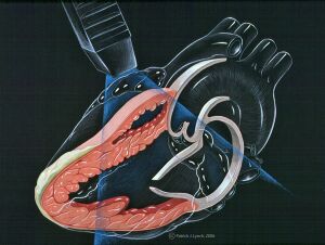
| A parasternal short axis on midpapillary level |
