Cause: Difference between revisions
No edit summary |
No edit summary |
||
| (3 intermediate revisions by 2 users not shown) | |||
| Line 5: | Line 5: | ||
{| class="wikitable" cellpadding="0" cellspacing="0" border="0" | {| class="wikitable" cellpadding="0" cellspacing="0" border="0" | ||
|[[Image: | |[[Image:Cause_flowchart.svg|600px]] | ||
|- | |- | ||
!Flowchart of the protocol CAUSE<cite>1</cite> | !Flowchart of the protocol CAUSE<cite>1</cite> | ||
| Line 47: | Line 47: | ||
{| class="wikitable" cellpadding="0" cellspacing="0" border="0" | {| class="wikitable" cellpadding="0" cellspacing="0" border="0" | ||
|[[Image:CAUSE02. | |[[Image:CAUSE02.svg|600px]] | ||
|- | |- | ||
!Ultrasound findings in a AP4Ch view. | !Ultrasound findings in a AP4Ch view. Adjusted from <cite>1</cite> | ||
|} | |} | ||
{| class="wikitable" cellpadding="0" cellspacing="0" border="0" | {| class="wikitable" cellpadding="0" cellspacing="0" border="0" width="600px" | ||
|Video | |Video | ||
|Video | |Video | ||
| Line 60: | Line 60: | ||
|} | |} | ||
==References== | ==References== | ||
<biblio> | <biblio> | ||
#1 pmid=17822831 | #1 pmid=17822831 | ||
</biblio> | </biblio> | ||
Latest revision as of 17:55, 13 April 2014
Cardiac Arrest Ultra Sound Exam (CAUSE) Cardiac arrest is a condition that often occurs in the Emergency Department, Intensive Care Unit and the surgical wards.
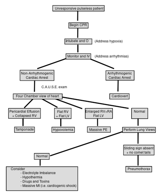
The use of ultrasound in resuscitation plays an important role in order to differentiate between the different causes of cardiac arrest, which are not a direct consequence of a primary ventricular arrhythmia. The most common and reversible causes of cardiac arrest include: severe hypovolemia, pneumothorax, cardiac tamponade, and massive pulmonary embolism. Identifying the underlying cause of cardiac arrest is one of the biggest challenges. This is the CAUSE protocol developed.

|
| Flowchart of the protocol CAUSE[1] |
|---|
Echocardiographic findings
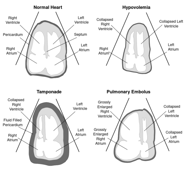
Echocardiography is the only mode with the possibility to respond in real-time during a cardiac arrest without interfering with the CPR. Moreover, direct diagnosis can be made when resuscitating thus saving time between the judgment and appropriate treatment. Below are several clinical findings that differentiate between cardiac tamponade, pneumothorax, pulmonary embolism, and severe hypovolemia secondary to abdominal aortic aneurysm.
| Subcostale views | Tamponade | Hypovolemie | Longembolie | Pneumothorax |
|---|---|---|---|---|
| VCI | >20mm | <5mm | >20mm | |
| Pericard | Many pericardial | "Sliding sign" | ||
| RV | Collapsed | Collapsed | Dilated | |
| LV | Flattened septum | Flattened septum |

|
| Ultrasound findings in a AP4Ch view. Adjusted from [1] |
|---|
| Video | Video |
| Tamponade | Pulmonary embolism[1] |
|---|