Normal Values of TEE
Jump to navigation
Jump to search
Reference values for normal adult TEE measurements
| Parameter | Mean ± SD (mm) | Range (mm) | |
|---|---|---|---|
| Right pulmonary artery diametera | 17 ± 3 | 12-22 | |
| Left upper pulmonary vein diameter | 11 ± 2 | 7-16 | |
| Left atrial appendage | Length | 28 ± 5 | 15-43 |
| Diameter | 16 ± 5 | 10-28 | |
| Superior vena cava diameter | 15 ± 3 | 8-20 | |
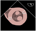
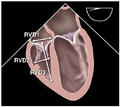
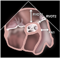
| Right ventricular outflow tract diameterb | 27 ± 4 | 16-36 | |
| Left atriumc | Anteroposterior diameter | 38 ± 6 | 20-52 |
| Medial-lateral diameter | 39 ± 7 | 24-52 | |
| Right atriumc | Anteroposterior diameter | 38 ± 5 | 28-52 |
| Medial-lateral diameter | 38 ± 6 | 29-53 | |
| Tricuspid annular diameterc | 28 ± 5 | 20-40 | |
| Mitral annular diameterc | 29 ± 4 | 20-38 | |
| Coronary sinus diameter | 6.6 ± 1.5 | 4-10 | |
| Left ventricled | Anteroposterior diameter (diastole) | 43 ± 7 | 33-55 |
| Medial-lateral diameter (diastole) | 42 ± 7 | 23-54 | |
| Anteroposterior diameter (systole) | 28 ± 6 | 18-40 | |
| Medial-lateral diameter (systole) | 27 ± 6 | 18-42 | |
| Aortic root diameterb | 28 ± 3 | 21-34 | |
| Descending thoracic aorta diameter | Proximal | 21 ± 4 | 14-30 |
| Distal | 20 ± 4 | 13-28 | |
| |||
Transesophageal Echocardiographic Anatomy
| ME two-chamber | Probe adjustment: neutral | Sector depth: ~14 cm |
|---|---|---|
| image | Primary diagnostic issues
|
Required structures
|
| ME LAX | Probe adjustment: neutral | Sector depth: ~12 cm |
| image | Primary diagnostic issues
|
Required structures
|
| ME mitral commissural | Probe adjustment: neutral | Sector depth: ~12 cm |
| image | Primary diagnostic issues
|
Required structures
|
| TG mid-SAX | Probe adjustment: neutral | Sector depth: ~12 cm |
| image | Primary diagnostic issues
|
Required structures
|
| TG two-chamber | Probe adjustment: neutral | Sector depth: ~12 cm |
| image | Primary diagnostic issues
|
Required structures
|
| TG RV inflow | Probe adjustment: neutral-rightward | Sector depth: ~12 cm |
| image | Primary diagnostic issues
|
Required structures
|
| TG RV inflow-outflow | Probe adjustment: neutral-rightward | Sector depth: ~14 cm |
| image | Primary diagnostic issues
|
Required structures
|
| TG basal SAX | Probe adjustment: neutral | Sector depth: ~12 cm |
| image | Primary diagnostic issues
|
Required structures
|
| TG LAX | Probe adjustment: neutral-leftward | Sector depth: ~12 cm |
| image | Primary diagnostic issues
|
Required structures
|
| Deep TG LAX | Probe adjustment: neutral | Sector depth: ~16 cm |
| image | Primary diagnostic issues
|
Required structures
|
| ||


