Parasternal short axis: Difference between revisions
Jump to navigation
Jump to search
No edit summary |
No edit summary |
||
| Line 4: | Line 4: | ||
{{IncludeFlash | {{IncludeFlash | ||
|flash_string=<flash>file=SAXPAP_normal.swf|quality=best|align=center|width=300|height=200</flash> | |flash_string=<flash>file=SAXPAP_normal.swf|quality=best|align=center|width=300|height=200</flash> | ||
|Description=A parasternal | |Description=A parasternal short axis on midpapillary level | ||
|file_name=SAXPAP_normal | |file_name=SAXPAP_normal | ||
}} | }} | ||
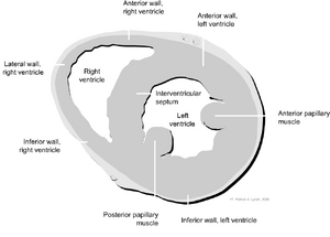
[[Image:Heart_normal_short_axis_echo.png|thumb|Image showing the left parasternal short axis transection (SAX) of the heart and the different structures]] | [[Image:Heart_normal_short_axis_echo.png|thumb|Image showing the left parasternal short axis transection (SAX) of the heart and the different structures]] | ||
{{clr}} | {{clr}} | ||
Revision as of 13:08, 20 September 2007
The parasternal short axis view (SAX) is usually made on three levels: Mitral valve levels, midpapillary, and apical level.
| <flash>file=SAXPAP_normal.swf |
| A parasternal short axis on midpapillary level |
