Repository: Difference between revisions
Jump to navigation
Jump to search
No edit summary |
Secretariat (talk | contribs) No edit summary |
||
| Line 36: | Line 36: | ||
|- | |- | ||
| rowspan="2" | | | rowspan="2" | | ||
| Right pulmonary artery diameter | | Right pulmonary artery diameter<sup>a</sup> | ||
| align="center" | 17 ± 3 | | align="center" | 17 ± 3 | ||
| align="center" | 12-22 | | align="center" | 12-22 | ||
| Line 58: | Line 58: | ||
| align="center" | 8-20 | | align="center" | 8-20 | ||
|- | |- | ||
| Right ventricular outflow tract diameter | | Right ventricular outflow tract diameter<sup>b</sup> | ||
| align="center" | 27 ± 4 | | align="center" | 27 ± 4 | ||
| align="center" | 16-36 | | align="center" | 16-36 | ||
|- | |- | ||
| rowspan="2" | Left atrium | | rowspan="2" | Left atrium<sup>c</sup> | ||
| Anteroposterior diameter | | Anteroposterior diameter | ||
| align="center" | 38 ± 6 | | align="center" | 38 ± 6 | ||
| Line 73: | Line 73: | ||
|- | |- | ||
| rowspan="5" | Right atrium | | rowspan="5" | Right atrium<sup>c</sup> | ||
| Anteroposterior diameter | | Anteroposterior diameter | ||
| align="center" | 38 ± 5 | | align="center" | 38 ± 5 | ||
| Line 82: | Line 82: | ||
| align="center" | 29-53 | | align="center" | 29-53 | ||
|- | |- | ||
| Tricuspid annular diameter | | Tricuspid annular diameter<sup>c</sup> | ||
| align="center" | 28 ± 5 | | align="center" | 28 ± 5 | ||
| align="center" | 20-40 | | align="center" | 20-40 | ||
|- | |- | ||
| Mitral annular diameter | | Mitral annular diameter<sup>c</sup> | ||
| align="center" | 29 ± 4 | | align="center" | 29 ± 4 | ||
| align="center" | 20-38 | | align="center" | 20-38 | ||
| Line 95: | Line 95: | ||
|- | |- | ||
| rowspan="5" | Left ventricle | | rowspan="5" | Left ventricle<sup>d</sup> | ||
| Anteroposterior diameter (diastole) | | Anteroposterior diameter (diastole) | ||
| align="center" | 43 ± 7 | | align="center" | 43 ± 7 | ||
| Line 112: | Line 112: | ||
| align="center" | 18-42 | | align="center" | 18-42 | ||
|- | |- | ||
| Aortic root diameter | | Aortic root diameter<sup>b</sup> | ||
| align="center" | 28 ± 3 | | align="center" | 28 ± 3 | ||
| align="center" | 21-34 | | align="center" | 21-34 | ||
| Line 129: | Line 129: | ||
| colspan="4" | | | colspan="4" | | ||
<ul> | <ul> | ||
<li><sup>a</sup> Right pulmonary artery diameter measured in midesophageal ascending aorta short-axis view.</li> | <li><sup>a</sup>Right pulmonary artery diameter measured in midesophageal ascending aorta short-axis view.</li> | ||
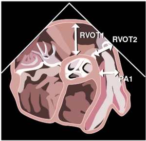
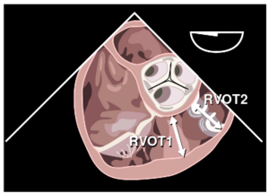
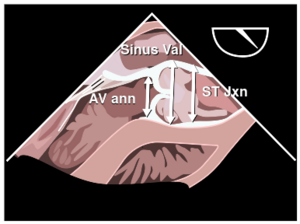
<li><sup>b</sup> Aortic root and right ventricular outflow tract diameters measured in the midesophageal right ventricular inflow/outflow tract view.</li> | <li><sup>b</sup>Aortic root and right ventricular outflow tract diameters measured in the midesophageal right ventricular inflow/outflow tract view.</li> | ||
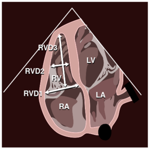
<li><sup>c</sup> Atrial (end-systole) and both mitral and tricuspid annular (mid-diastole) diameters measured in the midesophageal four-chamber view.</li> | <li><sup>c</sup>Atrial (end-systole) and both mitral and tricuspid annular (mid-diastole) diameters measured in the midesophageal four-chamber view.</li> | ||
<li><sup>d</sup> Left ventricular dimensions measured in transgastric mid short-axis view.</li> | <li><sup>d</sup>Left ventricular dimensions measured in transgastric mid short-axis view.</li> | ||
<li>SD, standard deviation.</li> | <li>SD, standard deviation.</li> | ||
<li>Adapted from Cohen G, White M, Sochowski R, et al. Reference values for normal transesophageal measurements. <em>J Am Soc Echocardiogr</em> 1995;8:221-230.</li> | <li>Adapted from Cohen G, White M, Sochowski R, et al. Reference values for normal transesophageal measurements. <em>J Am Soc Echocardiogr</em> 1995;8:221-230.</li> | ||
</ul> | </ul> | ||
|} | |} | ||
Revision as of 13:14, 26 June 2010
| <flash>file=MM0099.swf |
| gezondB |
| <flash>file=MM0100.swf |
| HF-B |
Reference values for normal adult TEE measurements
| Parameter | Mean ± SD (mm) | Range (mm) | |
|---|---|---|---|
| Right pulmonary artery diametera | 17 ± 3 | 12-22 | |
| Left upper pulmonary vein diameter | 11 ± 2 | 7-16 | |
| Left atrial appendage | Length | 28 ± 5 | 15-43 |
| Diameter | 16 ± 5 | 10-28 | |
| Superior vena cava diameter | 15 ± 3 | 8-20 | |
| Right ventricular outflow tract diameterb | 27 ± 4 | 16-36 | |
| Left atriumc | Anteroposterior diameter | 38 ± 6 | 20-52 |
| Medial-lateral diameter | 39 ± 7 | 24-52 | |
| Right atriumc | Anteroposterior diameter | 38 ± 5 | 28-52 |
| Medial-lateral diameter | 38 ± 6 | 29-53 | |
| Tricuspid annular diameterc | 28 ± 5 | 20-40 | |
| Mitral annular diameterc | 29 ± 4 | 20-38 | |
| Coronary sinus diameter | 6.6 ± 1.5 | 4-10 | |
| Left ventricled | Anteroposterior diameter (diastole) | 43 ± 7 | 33-55 |
| Medial-lateral diameter (diastole) | 42 ± 7 | 23-54 | |
| Anteroposterior diameter (systole) | 28 ± 6 | 18-40 | |
| Medial-lateral diameter (systole) | 27 ± 6 | 18-42 | |
| Aortic root diameterb | 28 ± 3 | 21-34 | |
| Descending thoracic aorta diameter | Proximal | 21 ± 4 | 14-30 |
| Distal | 20 ± 4 | 13-28 | |
| |||
