*[[Mmode echocardiography]]
*[[3D Echocardiography]]
223 bytes (22 words) - 17:22, 17 October 2023
Transthoracic echocardiography (TTE)
53 bytes (5 words) - 12:35, 14 September 2007
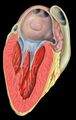
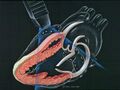
...erstood by every cardiologist and ultrasound technician. Three-dimensional echocardiography is also almost always the technique of choice because it rarely ahs any con
...hese waves form a pyramid-shaped ultrasound beam. With this technology, 3D echocardiography can be used to analyze and quantify anatomic abnormalities of the heart suc
3 KB (368 words) - 04:32, 15 June 2021
...eline of the American Society of Echoardiography / European Association of Echocardiography'''
* [http://ejechocard.oxfordjournals.org/content/11/2/202.full.pdf EAE Echocardiography in infective endocarditis]
2 KB (206 words) - 17:24, 17 October 2023
...mitral regurgitation. Transesophageal echocardiography with Doppler or 2D echocardiography and color flow Doppler imaging allows detailed assessment of the mitral val
1 KB (167 words) - 13:12, 13 March 2012
...le line of interrogation, similar to M-mode echocardiography. While M-mode echocardiography the intensity and location of the of a reflective spectral signal, in color
584 bytes (90 words) - 04:17, 11 March 2012
!Acute Echocardiography
!Stress Echocardiography
2 KB (185 words) - 15:39, 7 February 2014
 | File:Transesophageal echocardiography diagram.svg |Description = transesophageal echocardiography ultrasound diagram
[[Category:Cardiology]] [[Category:Echocardiography]] [[Category:Anatomical plates and drawings]] [[Category:Patrick Lynch]]
(524 × 555 (52 KB)) - 16:31, 17 October 2023 |
...nvasive investigations. Because in addition to improving quality contrast, echocardiography provides a secure and comprehensive investigation of cardiac structure, fun
Contrast echocardiography is used to enhance the echogenicity of blood (better signal/noise ratio) an
2 KB (249 words) - 13:40, 5 February 2014
 | File:Heart normal lpla echo.svg {{en|Heart normal left parasternal long axis echocardiography view}}
[[Category:Echocardiography]]
(483 × 379 (24 KB)) - 16:31, 17 October 2023 |
One of the earliest forms of cardiac ultrasound is Motion or “M”-mode echocardiography. Utilizing a rapid updating of more than 1,000 Hz, a single crystal rapidly
A great advantage of M-mode echocardiography is its high temporal resolution which makes possible the identification of
1 KB (180 words) - 14:09, 22 February 2012
...s segmental myocardial function by tracking natural acoustic markers in 2D echocardiography. The discrimination between transmurality states of myocardial infarction
402 bytes (50 words) - 03:15, 22 March 2012
*[https://techmed.sk/en/echo/new-examination/ Online free e-learning echocardiography (TECHmED)]
1 KB (146 words) - 08:13, 9 January 2021
This open access online echocardiography course and textbook for echocardiography is the second project of the [http://www.cardionetworks.org Cardionetworks
==History of Echocardiography==
6 KB (885 words) - 16:53, 21 February 2011
Echocardiography can identify the presence of mitral stenosis and determine its severity acc
863 bytes (126 words) - 12:48, 13 March 2012
 | File:TTE.jpeg |Description = Heart normal LPLA left parasternal long axis echocardiography view
(1,200 × 904 (1.23 MB)) - 16:31, 17 October 2023 |