Pulmonary: Difference between revisions
(Created page with "==Anatomy== The pulmonary valve is a tricuspid valve is similar in construction and size of the aortic valve. The valve has a right (R) -, a left (L) - and an anterior slip va...") |
|||
| (4 intermediate revisions by one other user not shown) | |||
| Line 1: | Line 1: | ||
==Anatomy== | ==Anatomy== | ||
The pulmonary valve is a tricuspid valve is similar in construction and size of the aortic valve. The valve has a right (R) -, a left (L) - and an anterior slip valve (A). The pulmonary valve is slightly above, left anterior aortic valve. | The pulmonary valve is a tricuspid valve is similar in construction and size of the aortic valve. The valve has a right (R) -, a left (L) - and an anterior slip valve (A). The pulmonary valve is slightly above, left anterior aortic valve.<cite>1</cite> | ||
[[Image:Pulmvalv. | [[Image:Pulmvalv.svg|400px]] | ||
==Echocardiographic views== | ==Echocardiographic views== | ||
| Line 16: | Line 16: | ||
==Stenosis== | ==Stenosis== | ||
Stenosis of the pulmonary valve is very rare . Although as with aortic stenosis , the cause may be located above or below the valve , the valve usually is a wrong engineered valve . Which is then | Stenosis of the pulmonary valve is very rare. Although as with aortic stenosis, the cause may be located above or below the valve, the valve usually is a wrong engineered valve. Which is then deformes into a dome formation with a small opening. | ||
{| class="wikitable" cellpadding="0" cellspacing="0" border="0" | {| class="wikitable" cellpadding="0" cellspacing="0" border="0" | ||
| Line 22: | Line 22: | ||
|colspan="2"|'''Causes''' <cite>2</cite> | |colspan="2"|'''Causes''' <cite>2</cite> | ||
|- | |- | ||
!Congenital | !width="20%"|Congenital | ||
|Tetralogy of Falot | |Tetralogy of Falot | ||
|- | |- | ||
| Line 32: | Line 32: | ||
==Tetralogy of Fallot== | ==Tetralogy of Fallot== | ||
Tetralogy of Fallot is a heart defect described by Etienne Fallot (1850-1911) , with four different heart defects occur: | Tetralogy of Fallot is a heart defect described by Etienne Fallot (1850-1911), with four different heart defects occur: | ||
*Pulmonaalstenose | *Pulmonaalstenose | ||
| Line 39: | Line 39: | ||
*About propelled aortic | *About propelled aortic | ||
Tetralogy of Fallot results in a lower concentration of oxygen in the blood by the mixing of oxygen - rich blood | Tetralogy of Fallot results in a lower concentration of oxygen in the blood by the mixing of oxygen - rich blood into the ventricles. The obstruction of the pulmonary valve ensures that the blood from the right ventricle to the aorta flows through the ventricular septal defect. Usually this syndrome is marked by cyanosis of the baby. Thus this disorder was formerly called "blue baby syndrome". There are also "pink fallots", where the obstruction of the pulmonary valve is less severe. These patients are usually detected with an image of heart failure by excessive flow of the pulmonary vascular tree. In rare cases, there is a balanced position, wherein the stenosis is large enough in order to protect (against over-flow), and is low enough not to cause too much cyanosis pulmonary vascular. | ||
Click [http://www.tetralogievanfallot.nl/|'''here'''] for more info on Tetralogy of Fallot | Click [http://www.tetralogievanfallot.nl/|'''here'''] for more info on Tetralogy of Fallot | ||
==Insufficiency== | ==Insufficiency== | ||
Pulmonary insufficiency is a volume load on the RV. Important insufficiency will lead to RV dilation. This volume will load the RV into long endure, but will eventually lead to RV failure. | |||
{| class="wikitable" cellpadding="0" cellspacing="0" border="0" | {| class="wikitable" cellpadding="0" cellspacing="0" border="0" | ||
| Line 50: | Line 50: | ||
|colspan="2"|'''Causes''' <cite>3</cite> | |colspan="2"|'''Causes''' <cite>3</cite> | ||
|- | |- | ||
!Physiologic | !width="20%"|Physiologic | ||
|Is found in 40-80% of people | |Is found in 40-80% of people | ||
|- | |- | ||
!Congenital | !Congenital | ||
| | |Incorrectly laid valve cusps or absence of (partial) cover slip. | ||
|- | |- | ||
!valign="top"|Acquired | !valign="top"|Acquired | ||
| | | | ||
*As a result of pulmonary hypertension. | *As a result of pulmonary hypertension. | ||
*By balloon dilation or plastic PV at correcting | *By balloon dilation or plastic PV at correcting pulmonary stenosis. | ||
*Carcinoid | *Carcinoid | ||
*Endocarditis. | *Endocarditis. | ||
| Line 66: | Line 66: | ||
==References== | ==References== | ||
<biblio> | <biblio> | ||
#1 [ | #1 [http://www.geisinger.kramesonline.com/3,S,89116 Geisinger - How the Heart Works] | ||
#2 pmid=19065003 | #2 pmid=19065003 | ||
#3 pmid=20375260 | #3 pmid=20375260 | ||
</biblio> | </biblio> | ||
Latest revision as of 09:31, 29 March 2014
Anatomy
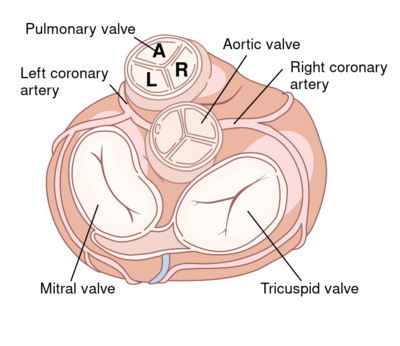
The pulmonary valve is a tricuspid valve is similar in construction and size of the aortic valve. The valve has a right (R) -, a left (L) - and an anterior slip valve (A). The pulmonary valve is slightly above, left anterior aortic valve.1
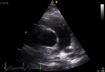
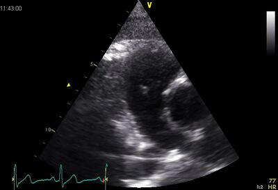
Echocardiographic views

|

|
| PSAX ao | Plax by tilted |
|---|
Stenosis
Stenosis of the pulmonary valve is very rare. Although as with aortic stenosis, the cause may be located above or below the valve, the valve usually is a wrong engineered valve. Which is then deformes into a dome formation with a small opening.
| Causes 2 | |
| Congenital | Tetralogy of Falot |
|---|---|
| Acquired |
|
Tetralogy of Fallot
Tetralogy of Fallot is a heart defect described by Etienne Fallot (1850-1911), with four different heart defects occur:
- Pulmonaalstenose
- VSD
- RV Hypertrophy
- About propelled aortic
Tetralogy of Fallot results in a lower concentration of oxygen in the blood by the mixing of oxygen - rich blood into the ventricles. The obstruction of the pulmonary valve ensures that the blood from the right ventricle to the aorta flows through the ventricular septal defect. Usually this syndrome is marked by cyanosis of the baby. Thus this disorder was formerly called "blue baby syndrome". There are also "pink fallots", where the obstruction of the pulmonary valve is less severe. These patients are usually detected with an image of heart failure by excessive flow of the pulmonary vascular tree. In rare cases, there is a balanced position, wherein the stenosis is large enough in order to protect (against over-flow), and is low enough not to cause too much cyanosis pulmonary vascular.
Click here for more info on Tetralogy of Fallot
Insufficiency
Pulmonary insufficiency is a volume load on the RV. Important insufficiency will lead to RV dilation. This volume will load the RV into long endure, but will eventually lead to RV failure.
| Causes 3 | |
| Physiologic | Is found in 40-80% of people |
|---|---|
| Congenital | Incorrectly laid valve cusps or absence of (partial) cover slip. |
| Acquired |
|
References
<biblio>
- 1 Geisinger - How the Heart Works
- 2 pmid=19065003
- 3 pmid=20375260
</biblio>
