Case 36 and Case 34: Difference between pages
(Difference between pages)
Jump to navigation
Jump to search
Secretariat (talk | contribs) (Created page with '{{EchoCase |Title = Thrombus of prosthetic aortic valve |CasePresentation = |Courtesy = AMC Echolab, AMC, The Netherlands |filepointer1=<flash>file=E00266.swf|quality=best|a...') |
No edit summary |
||
| Line 1: | Line 1: | ||
{{EchoCase | {{EchoCase | ||
|Title = | |Title = Endocarditis of aortic valve | ||
|CasePresentation = | |CasePresentation = | ||
|Courtesy = [[AMC Echolab, AMC, The Netherlands]] | |Courtesy = [[AMC Echolab, AMC, The Netherlands]] | ||
|filepointer1=< | |filepointer1=<html5media height="350" width="279" autoplay="true" loop="true">File:E00252.mp4</html5media> | ||
|file_name1= | |file_name1=E00252 | ||
|descriptionfile1= | |descriptionfile1=PLAX: vegetations on aortic valve, dilated left ventricle and left atrium | ||
|filepointer2=< | |filepointer2=<html5media height="350" width="279" autoplay="true" loop="true">File:E00253.mp4</html5media> | ||
|file_name2= | |file_name2=E00253 | ||
|descriptionfile2= | |descriptionfile2=PLAX: Color Doppler, severe aortic regurgitation, excentric jet, and moderate mitral regurgitation | ||
|filepointer3=[[File: | |filepointer3=<html5media height="350" width="279" autoplay="true" loop="true">File:E00254.mp4</html5media> | ||
| | |file_name3=E00254 | ||
| | |descriptionfile3=PSAX: thickend aortic valve | ||
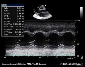
|filepointer4=[[File:E00255.jpg|350px|left]] | |||
|file_name4= | |||
|descriptionfile4=M-Mode: dilated left ventricle | |||
|filepointer5=<html5media height="350" width="279" autoplay="true" loop="true">File:E00256.mp4</html5media> | |||
|file_name5=E00256 | |||
|descriptionfile5=A3CH: Color Doppler, severe aortic regurgitation, excentric jet | |||
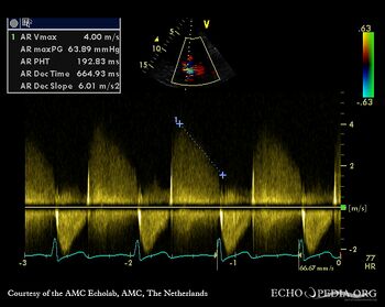
|filepointer6=[[File:E00257.jpg|350px|left]] | |||
|file_name6= | |||
|descriptionfile6=Continuous-wave signal of severe aortic regurgitation | |||
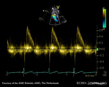
|filepointer7=[[File:E00258.jpg|350px|left]] | |||
|file_name7= | |||
|descriptionfile7=Subcostal view: pulsed-wave Doppler signal from abdominal aorta: diastolic flow reversal | |||
}} | }} | ||
Revision as of 19:26, 20 February 2016
| Courtesy of: AMC Echolab, AMC, The Netherlands | |
| <html5media height="350" width="279" autoplay="true" loop="true">File:E00252.mp4</html5media> | <html5media height="350" width="279" autoplay="true" loop="true">File:E00253.mp4</html5media> |
| PLAX: vegetations on aortic valve, dilated left ventricle and left atrium | PLAX: Color Doppler, severe aortic regurgitation, excentric jet, and moderate mitral regurgitation |
| <html5media height="350" width="279" autoplay="true" loop="true">File:E00254.mp4</html5media> | |
| PSAX: thickend aortic valve | M-Mode: dilated left ventricle |
| <html5media height="350" width="279" autoplay="true" loop="true">File:E00256.mp4</html5media> | |
| A3CH: Color Doppler, severe aortic regurgitation, excentric jet | Continuous-wave signal of severe aortic regurgitation |
| Subcostal view: pulsed-wave Doppler signal from abdominal aorta: diastolic flow reversal | |


