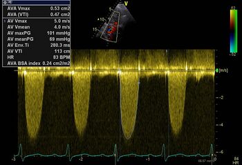
Aortic Valve Stenosis
Aortic stenosis quantification
| Slight | Moderate | Severe | |
|---|---|---|---|
| PG max | 15-40 mmHg | 40-70 mmHg | 70-100 mmHg (> 100 mmHg severe AoS) |
| PG mean | <20mmHg | 20-40mmHg> | 40mmHg |
| AVA> | 1.5cm2 | 1.5-1cm2 | <1cm2 |
| Dimensionless index> | 0.50 | 0.25 to 0.50 | <0.25 |
| Vmax | <3m/s | 3-4m/s | >4m/s |
| AVA BSAindex | >0.85cm2/m2 | 0.85-0.60cm2/m2 | <0.60cm2/m2 |

|
|
| Serious AoS: PG max 101mmHg, PG mean 69mmHg[1] | PHT moderate Aol [1] |
|---|---|
| Click here to calculate AGM | |
Aortic valve area (AVA)
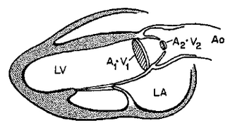
For the calculation of the aortic valve surface, the continuity equation can be applied.
Dimensionless index (DI)
If you are unable to get a good measurement of the LVOT can also the ratio of the velocity in the LVOT and the speed of the aortic valve to give a good impression about the severity of aortic stenosis .
DI = V LVOT/V aorta In a DI = 25, there is almost always a severe aortic valve stenosis .
Low- gradient aortic stenosis vs . pseudo stenosis
In an aortic valve stenosis with a low gradient (<30 mmHg ), and poor left ventricular function is to be a valve surface calculated . If an AVA <1 cm ² is calculated can be a severe aortic valve stenosis are present despite the low gradient . However, it could also be that the valve does not open properly due to low cardiac output ( pseudo stenosis ) . To gain more insight into the aortic valve or a "true " or stenosis has a "pseudo stenosis " seems dobutamine stress worthwhile . If during dobutamine infusion up to 20 micrograms / kg there is an increase of more than 20 % in the LVOT VTI (ie cardiac output ) and the calculated valve area remains below 1 cm ² then there is a true aortic stenosis . If not , and there appears to be a contractile reserve (which is predictive of a better prognosis after aortic valve replacement ) it is called a pseudo stenosis.
