Cardiomyopathy: Difference between revisions
Jump to navigation
Jump to search
No edit summary |
No edit summary |
||
| (7 intermediate revisions by one other user not shown) | |||
| Line 22: | Line 22: | ||
|} | |} | ||
Click [http://www.cardiogenetica.nl/index.php?menuID=2 '''here'''] for detailed information on various cardiomyopathy | Click [http://www.cardiogenetica.nl/index.php?menuID=2 '''here'''] for detailed information on various cardiomyopathy. | ||
{| class="wikitable" cellpadding="0" cellspacing="0" border="0" | {| class="wikitable" cellpadding="0" cellspacing="0" border="0" | ||
|+ Listed below are the main disorders and their characteristics with examples.<cite>1</cite> | |||
|- | |- | ||
!Condition | !Condition | ||
!Features | !Features | ||
!Example | !Example | ||
| Line 32: | Line 33: | ||
!rowspan="4" valign="top"|Arrythmogene right ventricular cardiomyopathy (ARVC) | !rowspan="4" valign="top"|Arrythmogene right ventricular cardiomyopathy (ARVC) | ||
|rowspan="4" valign="top"| | |rowspan="4" valign="top"| | ||
*Fibrofatty degeneration of the RV . | *Fibrofatty degeneration of the RV. | ||
*Myocardial degeneration leads to RV dilation and poor RVF . | *Myocardial degeneration leads to RV dilation and poor RVF. | ||
*Ventricular fibrillation by slow conduction velocities , guide block and spatial variation in conduction velocity . | *Ventricular fibrillation by slow conduction velocities, guide block and spatial variation in conduction velocity. | ||
*Aneurysms of the RV free wall . | *Aneurysms of the RV free wall. | ||
* | *Echodense moderator band and myocardial RV free wall. | ||
*Genetic component | *Genetic component | ||
*Rare 1 : 5000 people. | *Rare 1:5000 people. | ||
|Video | |Video | ||
|- | |- | ||
| Line 47: | Line 48: | ||
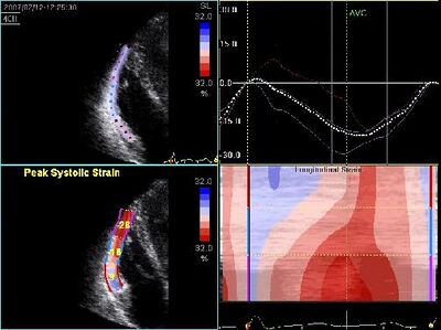
!Decreased RV strain in ARVC | !Decreased RV strain in ARVC | ||
|- | |- | ||
!rowspan=" | !rowspan="6" valign="top"|Dilated cardiomyopathy (DCM) | ||
|rowspan=" | |rowspan="6" valign="top"| | ||
*It is the most common form of cardiomyopathy. | |||
Poor LVF and LV dilatation . | *Also known as congestive cardiomyopathy. | ||
Arrhythmias ( atrial fibrillation 20-30 % ) . | *Poor LVF and LV dilatation. | ||
Clot formation, which may lead to thrombo -embolic complications. | *Arrhythmias (atrial fibrillation 20-30%). | ||
Often accompanied by pulmonary hypertension , dilation of other compartments , and an insufficiency of mitral and / or tricuspid valve | *Clot formation, which may lead to thrombo-embolic complications. | ||
Familial DCM 's common to autosomal dominant, autosomal recessive and sex-linked inheritance . | *Often accompanied by pulmonary hypertension, dilation of other compartments, and an insufficiency of mitral and/or tricuspid valve | ||
*Familial DCM's common to autosomal dominant, autosomal recessive and sex-linked inheritance. | |||
( post - ) infectious : various viruses and bacteria , as the final stage of myocarditis. | *Causes: | ||
intoxication : cocaine, alcohol abuse . | **(post-) infectious: various viruses and bacteria, as at the final stage of myocarditis. | ||
iatrogenic : some chemostatica , X-ray radiation . | **intoxication: cocaine, alcohol abuse. | ||
Metabolic : vitamin B1 deficiency . | **iatrogenic: some chemostatica, X-ray radiation. | ||
- idiopathic : In approximately 30 % of cases, no cause is found . | **Metabolic: vitamin B1 deficiency. | ||
**-idiopathic: In approximately 30% of cases, no cause is found | |||
Dilated LV on AP4CH | |[[Image:DCM01.jpg|400px]] | ||
|- | |||
Dilated LV on | !Dilated LV on AP4CH | ||
|- | |||
EPSS is a useful measurement | |[[Image:LVF slecht05.jpg|400px]] | ||
Hypertrophic cardiomyopathy ( HCM ) | |- | ||
!Dilated LV on PLAX | |||
65% | |- | ||
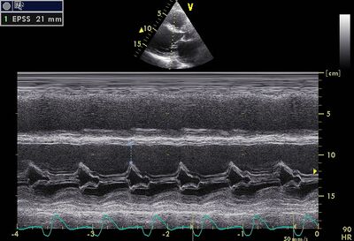
35 % | |[[Image:EPSS01.jpg|400px]] | ||
Small LV lumen . | |- | ||
Preserved systolic LV function ( EF normal or slightly decreased) | !EPSS is a useful measurement to follow up DCM | ||
Diastolic dysfunction . | |- | ||
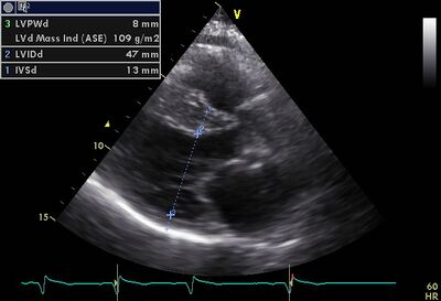
Autosomal dominant progressive deviation from nature . | !rowspan="6" valign="top"|Hypertrophic cardiomyopathy (HCM) | ||
|rowspan="6" valign="top"| | |||
Heart failure can be caused by the rigidity of the thickened heart muscle ( diastolic heart failure ) , by an obstruction in the LVOT (SAM ) is associated with mitral valvular insufficiency . The course of the disease is progressive . | *65% asymmetric hypertrophy of the myocardium, usually ventricular septum sometimes apical involvement. | ||
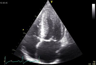
Occurs in persons 1:500-1000 | *35% symmetrical hypertrophy of the myocardium (not to be confused with aortic stenosis or hypertension). | ||
*Small LV lumen. | |||
*Preserved systolic LV function (EF normal or slightly decreased) | |||
*Diastolic dysfunction. | |||
*Autosomal dominant progressive deviation from nature. | |||
*Could be associated with sudden cardiac death due to ventricular fibrillation, an increased risk of thromboembolism. | |||
*Heart failure can be caused by the rigidity of the thickened heart muscle (diastolic heart failure), by an obstruction in the LVOT (SAM) and is associated with mitral valvular insufficiency. The course of the disease is progressive. | |||
Non - compaction cardiomyopathy ( NCCMP ) | *Occurs in persons 1:500-1000 | ||
|[[Image:Asym.cmp1.jpg|400px]] | |||
|- | |||
Jenni criteria ( Heart 2007) . | !Asymmetric hypertrophy | ||
Also called insulated non compaction of the ventricular myocardium ( INVM ) , is a rare form of congenital heart disease in which the tissue of the ventricular myocardium is constructed in terms of texture | |- | ||
|[[Image:HCM01.jpg|400px]] | |||
It is a congenital defect , which occurs in the 20th week of pregnancy . | |- | ||
The condition is expressed by heart failure, arrhythmias, and an increased risk of thrombus formation. | !Symmetrical hypertrophy | ||
The disorder often manifests itself later in life and has a high mortality rate due to heart failure and arrhythmias . | |- | ||
|[[Image:ApicHCM.jpg|400px]] | |||
NCCMP with crypts and apical midventriculair | |- | ||
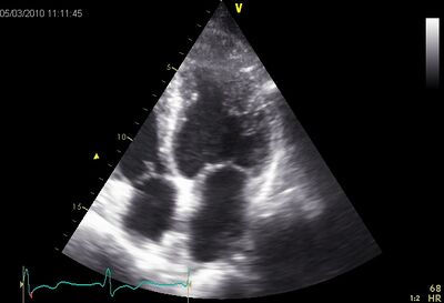
!Apical hypertrophy | |||
NCCCMP on PSax | |- | ||
!rowspan="4" valign="top"|Non-compaction cardiomyopathy (NCCMP) | |||
|rowspan="4" valign="top"| | |||
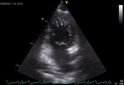
*LV wall has a spongy appearance. | |||
Stiffened myocardium. | *Jenni criteria (Heart 2007). | ||
This form comes after at least 1 and is usually associated with storage diseases , such as sarcoidosis , amyloidosis , and the like. | *Also called insulated non compaction of the ventricular myocardium (INVM), it is a rare form of congenital heart disease in which the tissue of the ventricular myocardium is not well constructed in terms of texture. | ||
Preserved systolic LVF . LV is not dilated . | *After HCM DCM, it is the most common cause of primary cardiomyopathy in children. | ||
Diastolic dysfunction ( see fig.) | *It is a congenital defect, which occurs in the 20th week of pregnancy. | ||
LV and RV may be hypertrophied . | *The condition is expressed by heart failure, arrhythmias, and an increased risk of thrombus formation. | ||
Dilated atria and VCI . | *The disorder often manifests itself later in life and has a high mortality rate due to heart failure and arrhythmias. | ||
Pulmonary hypertension . | |[[Image:NCCMP02.jpg|400px]] | ||
Myocardial echo during and amyloidosis speckled | |- | ||
!NCCMP with crypts and apical midventriculair | |||
Abnormally low É in restrictive | |- | ||
Tako- tsubo cardiomyopathy | |Video | ||
|- | |||
Apical ballooning , | !NCCCMP on PSax | ||
|- | |||
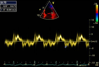
Is more common in women than in men , the average age of 62 to 75 years . | !rowspan="2" valign="top"|Restrictive cardiomyopathy | ||
|rowspan="2" valign="top"| | |||
Also called "broken heart syndrome" or " Stress CMP " | *Stiffened myocardium. | ||
LV | *This form comes after at least 1 and is usually associated with storage diseases, such as sarcoidosis, amyloidosis, and the like. | ||
*Preserved systolic LVF. LV is not dilated. | |||
Apical ballooning | *Diastolic dysfunction (see fig.) | ||
*LV and RV may be hypertrophied. | |||
*Dilated atria and VCI. | |||
*Pulmonary hypertension. | |||
*Myocardial echo during and amyloidosis speckled | |||
|[[Image:Restrcmp.jpg|400px]] | |||
|- | |||
!Abnormally low É in restrictive cardiomyopathy | |||
|- | |||
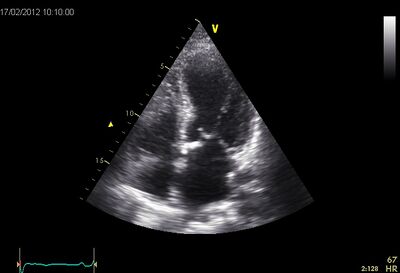
!rowspan="2" valign="top"|Tako-tsubo cardiomyopathy | |||
|rowspan="2" valign="top"| | |||
*Takotsubo is named after the ceramic pots used to trap octopus in Japan. | |||
*Apical ballooning, akinetic of the apex. This gives the LV the octupus trap shape. | |||
*Manifests itself as an acute myocardial infarction with ST elevations, however, no significant coronary artery disease. | |||
*Is more common in women than in men, the average age of 62 to 75 years. | |||
*Stress induced, is triggered by an acute illness or intense emotional or physical stress | |||
*Also called "broken heart syndrome" or "Stress CMP". | |||
*LV normalizes in a few days to several weeks. | |||
|[[Image:TakoTsubo01.jpg|400px]] | |||
|- | |||
!Apical ballooning | |||
|} | |} | ||
==References== | ==References== | ||
<biblio> | <biblio> | ||
#1 | #1 [http://www.uptodate.com/contents/echocardiographic-recognition-of-cardiomyopathies?source=search_result&search=cardiomyopathy+echo&selectedTitle=1~150| Echocardiographic | ||
#2 recognition of cardiomyopathies] | |||
#3 [https://vetocorleone.com/download-idn-poker/ Download IDN Poker APk] | |||
#4 [https://vetocorleone.com/deposit-idn-poker/ Deposit IDNPoker Terbaru] | |||
#5 [https://vetocorleone.com/daftar-idn-poker/ Daftar IDN Poker Online] | |||
#6 [https://vetocorleone.com/ IDN Poker] | |||
#7 [https://daftar-idn-poker88.wildapricot.org/ IDN Poker] | |||
#8 [https://togelonlineku3.wildapricot.org/ Togel Hongkong] | |||
</biblio> | </biblio> | ||
Latest revision as of 04:33, 15 June 2021
Cardiomyopathy (CMP) is a collective term for various diseases of the heart muscle (myocardium). For various reasons, the function of the myocardium decreased (see table). The different variants of a CMP are generally classified on the basis of echocardiographic characteristics.
| LV function decline in most common cardiomyopathy | ||
|---|---|---|
| Systolic function | Diastolic Function | |
| Dilated CMP | ↓ | =/↓ |
| Hypertrophic CMP | ↑ | ↓ |
| Restrictive CMP | = | ↓ |
Click here for detailed information on various cardiomyopathy.
| Condition | Features | Example |
|---|---|---|
| Arrythmogene right ventricular cardiomyopathy (ARVC) |
|
Video |
| Echodense RV free wall for suspected ARVC | ||
| ||
| Decreased RV strain in ARVC | ||
| Dilated cardiomyopathy (DCM) |
|
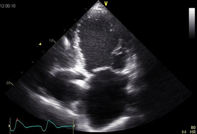
|
| Dilated LV on AP4CH | ||
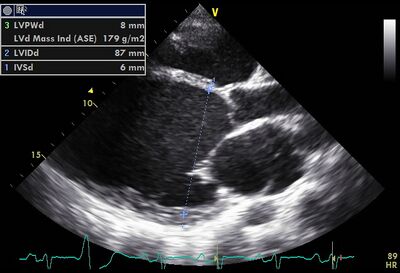
| ||
| Dilated LV on PLAX | ||
| ||
| EPSS is a useful measurement to follow up DCM | ||
| Hypertrophic cardiomyopathy (HCM) |
|
|
| Asymmetric hypertrophy | ||
| ||
| Symmetrical hypertrophy | ||
| ||
| Apical hypertrophy | ||
| Non-compaction cardiomyopathy (NCCMP) |
|
|
| NCCMP with crypts and apical midventriculair | ||
| Video | ||
| NCCCMP on PSax | ||
| Restrictive cardiomyopathy |
|
|
| Abnormally low É in restrictive cardiomyopathy | ||
| Tako-tsubo cardiomyopathy |
|
|
| Apical ballooning |
References
-
recognition of cardiomyopathies]