Case 153: Difference between revisions
Jump to navigation
Jump to search
Secretariat (talk | contribs) (Created page with '{{EchoCase |Title = Aortic dissection type A |CasePresentation = |Courtesy = AMC Echolab, AMC, The Netherlands |filepointer1=<flash>file=E00791.swf|quality=best|align=center...') |
m (Replace html5media with gif) |
||
| (One intermediate revision by one other user not shown) | |||
| Line 4: | Line 4: | ||
|Courtesy = [[AMC Echolab, AMC, The Netherlands]] | |Courtesy = [[AMC Echolab, AMC, The Netherlands]] | ||
|filepointer1= | |filepointer1=[[File:E00791.gif|350px]] | ||
|file_name1=E00791 | |file_name1=E00791 | ||
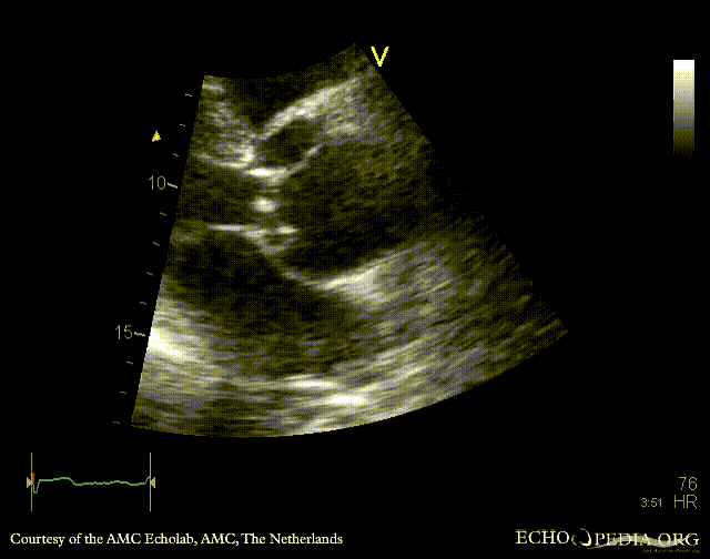
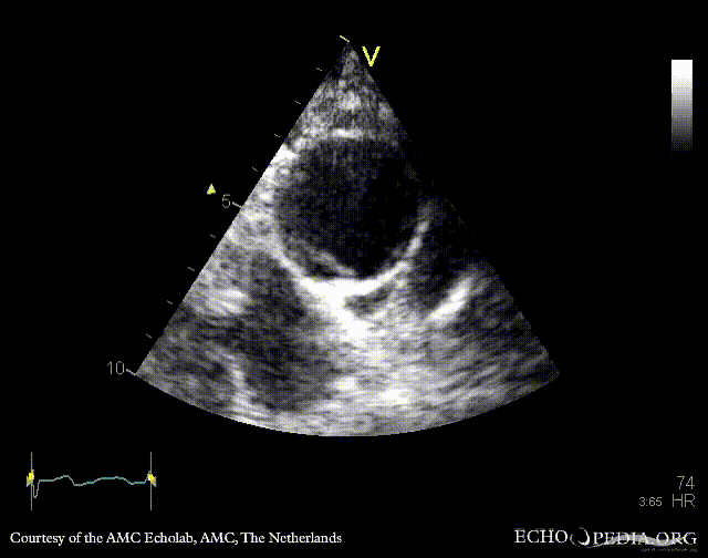
|descriptionfile1=PLAX: dilated aortic root, dissection flap in ascending aorta | |descriptionfile1=PLAX: dilated aortic root, dissection flap in ascending aorta | ||
| Line 12: | Line 12: | ||
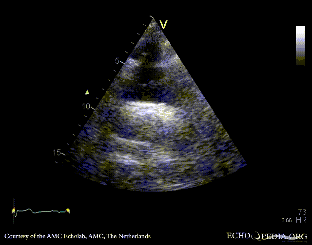
|descriptionfile2=PLAX: diameter of aortic root | |descriptionfile2=PLAX: diameter of aortic root | ||
|filepointer3= | |filepointer3=[[File:E00793.gif|350px]] | ||
|file_name3=E00793 | |file_name3=E00793 | ||
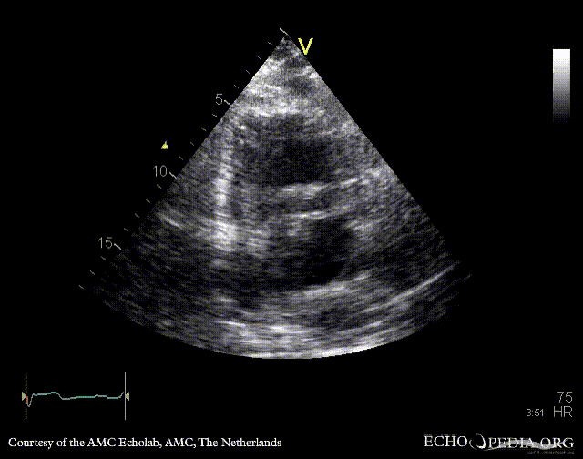
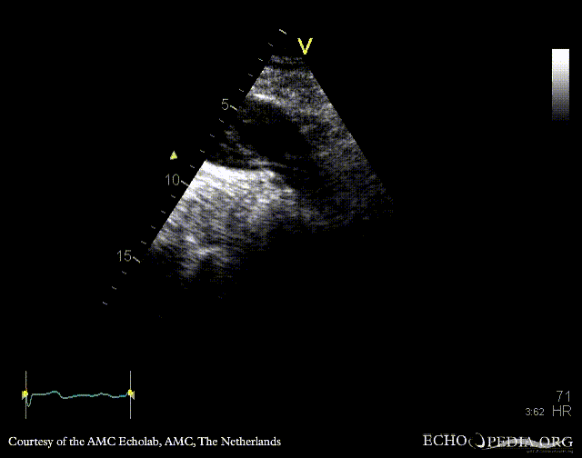
|descriptionfile3=PSAX | |descriptionfile3=PSAX | ||
|filepointer4= | |filepointer4=[[File:E00794.gif|350px]] | ||
|file_name4=E00794 | |file_name4=E00794 | ||
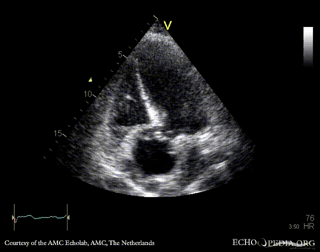
|descriptionfile4=A5CH: dilated aortic root, dissection flap in ascending aorta | |descriptionfile4=A5CH: dilated aortic root, dissection flap in ascending aorta | ||
|filepointer5= | |filepointer5=[[File:E00795.gif|350px]] | ||
|file_name5=E00795 | |file_name5=E00795 | ||
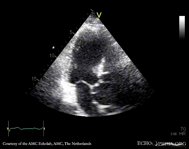
|descriptionfile5=A3CH | |descriptionfile5=A3CH | ||
|filepointer6= | |filepointer6=[[File:E00796.gif|350px]] | ||
|file_name6=E00796 | |file_name6=E00796 | ||
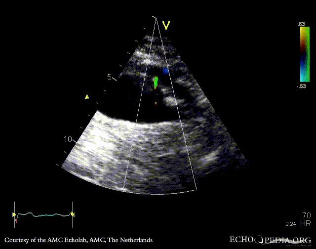
|descriptionfile6=A3CH with Color Doppler: mild aortic regurgitation | |descriptionfile6=A3CH with Color Doppler: mild aortic regurgitation | ||
|filepointer7= | |filepointer7=[[File:E00797.gif|350px]] | ||
|file_name7=E00797 | |file_name7=E00797 | ||
|descriptionfile7=Suprasternal view: dissection flap in aortic arch | |descriptionfile7=Suprasternal view: dissection flap in aortic arch | ||
|filepointer8= | |filepointer8=[[File:E00798.gif|350px]] | ||
|file_name8=E00798 | |file_name8=E00798 | ||
|descriptionfile8=Subcostal view: dissection flap in abdominal aorta | |descriptionfile8=Subcostal view: dissection flap in abdominal aorta | ||
|filepointer9= | |filepointer9=[[File:E00799.gif|350px]] | ||
|file_name9=E00799 | |file_name9=E00799 | ||
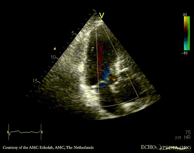
|descriptionfile9=Subcostal view with Color Doppler: flow between true and false lumen | |descriptionfile9=Subcostal view with Color Doppler: flow between true and false lumen | ||
|filepointer10= | |filepointer10=[[File:E00800.gif|350px]] | ||
|file_name10=E00800 | |file_name10=E00800 | ||
|descriptionfile10=Suprasternal view: dissection flap in ascending aorta | |descriptionfile10=Suprasternal view: dissection flap in ascending aorta | ||
|filepointer11= | |filepointer11=[[File:E00801.gif|350px]] | ||
|file_name11=E00801 | |file_name11=E00801 | ||
|descriptionfile11=Suprasternal view: dissection flap in ascending aorta | |descriptionfile11=Suprasternal view: dissection flap in ascending aorta | ||
}} | }} | ||
Latest revision as of 20:02, 1 December 2023
| Courtesy of: AMC Echolab, AMC, The Netherlands | |

|
 |
| PLAX: dilated aortic root, dissection flap in ascending aorta | PLAX: diameter of aortic root |

|

|
| PSAX | A5CH: dilated aortic root, dissection flap in ascending aorta |

|

|
| A3CH | A3CH with Color Doppler: mild aortic regurgitation |

|

|
| Suprasternal view: dissection flap in aortic arch | Subcostal view: dissection flap in abdominal aorta |

|

|
| Subcostal view with Color Doppler: flow between true and false lumen | Suprasternal view: dissection flap in ascending aorta |