Case 158: Difference between revisions
Jump to navigation
Jump to search
No edit summary |
m (Replace html5media with gif) |
||
| Line 4: | Line 4: | ||
|Courtesy = [[AMC Echolab, AMC, The Netherlands]] | |Courtesy = [[AMC Echolab, AMC, The Netherlands]] | ||
|filepointer1= | |filepointer1=[[File:E00832.gif|350px]] | ||
|file_name1=E00832 | |file_name1=E00832 | ||
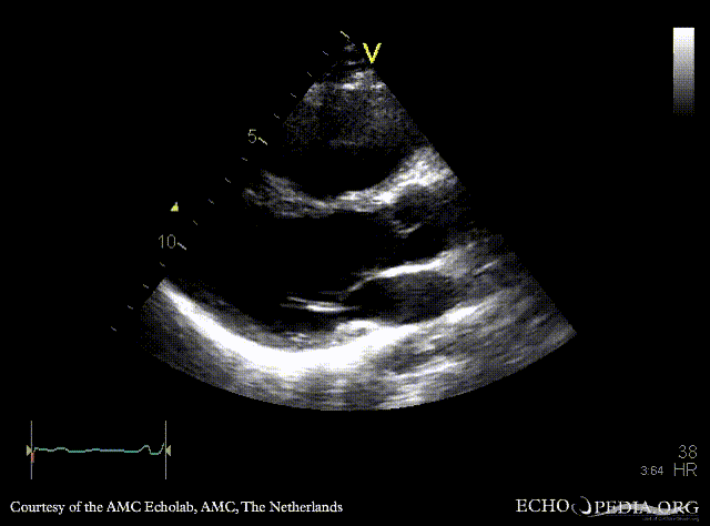
|descriptionfile1=PLAX: large perimembraneous ventricular septum defect | |descriptionfile1=PLAX: large perimembraneous ventricular septum defect | ||
|filepointer2= | |filepointer2=[[File:E00833.gif|350px]] | ||
|file_name2=E00833 | |file_name2=E00833 | ||
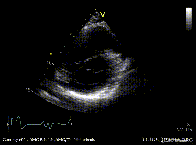
|descriptionfile2=PSAX: dilated right ventricle, flattening of interventricular septum | |descriptionfile2=PSAX: dilated right ventricle, flattening of interventricular septum | ||
|filepointer3= | |filepointer3=[[File:E00834.gif|350px]] | ||
|file_name3=E00834 | |file_name3=E00834 | ||
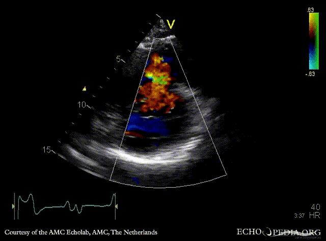
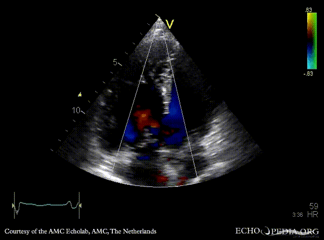
|descriptionfile3=PSAX with Color Doppler: right to left shunt (syndrome of Eisenmenger) | |descriptionfile3=PSAX with Color Doppler: right to left shunt (syndrome of Eisenmenger) | ||
|filepointer4= | |filepointer4=[[File:E00835.gif|350px]] | ||
|file_name4=E00835 | |file_name4=E00835 | ||
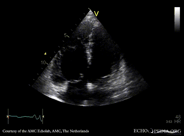
|descriptionfile4=A4CH: dilated right ventricle, poor function, VSD | |descriptionfile4=A4CH: dilated right ventricle, poor function, VSD | ||
|filepointer5= | |filepointer5=[[File:E00836.gif|350px]] | ||
|file_name5=E00836 | |file_name5=E00836 | ||
|descriptionfile5=A4CH with Color Doppler: right to left shunt | |descriptionfile5=A4CH with Color Doppler: right to left shunt | ||
Latest revision as of 20:05, 1 December 2023
| Courtesy of: AMC Echolab, AMC, The Netherlands | |

|

|
| PLAX: large perimembraneous ventricular septum defect | PSAX: dilated right ventricle, flattening of interventricular septum |

|

|
| PSAX with Color Doppler: right to left shunt (syndrome of Eisenmenger) | A4CH: dilated right ventricle, poor function, VSD |

|
 |
| A4CH with Color Doppler: right to left shunt | Continuous-wave Doppler signal of tricuspid regurgitation: severe pulmonary hypertension |