Case 30: Difference between revisions
Jump to navigation
Jump to search
No edit summary |
m (Replace html5media with gif) |
||
| (3 intermediate revisions by one other user not shown) | |||
| Line 4: | Line 4: | ||
|Courtesy = [[AMC Echolab, AMC, The Netherlands]] | |Courtesy = [[AMC Echolab, AMC, The Netherlands]] | ||
|filepointer1= | |filepointer1=[[File:E00243.gif|350px]] | ||
|file_name1=E00243 | |file_name1=E00243 | ||
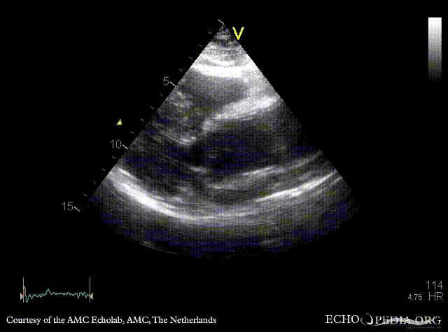
|descriptionfile1=PLAX: dilated aortic root and ascending aorta | |descriptionfile1=PLAX: dilated aortic root and ascending aorta | ||
|filepointer2= | |filepointer2=[[File:E00244.gif|350px]] | ||
|file_name2=E00244 | |file_name2=E00244 | ||
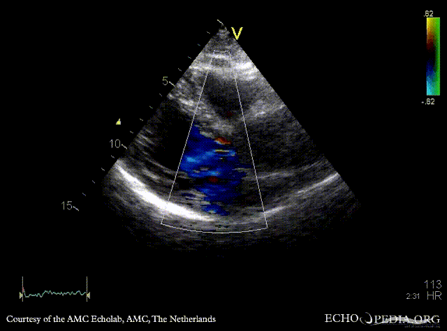
|descriptionfile2=PLAX: Color Doppler, mild aortic regurgitation | |descriptionfile2=PLAX: Color Doppler, mild aortic regurgitation | ||
|filepointer3= | |filepointer3=[[File:E00245.gif|350px]] | ||
|file_name3=E00245 | |file_name3=E00245 | ||
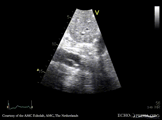
|descriptionfile3=Subcostal view: dissection flap in abdominal aorta | |descriptionfile3=Subcostal view: dissection flap in abdominal aorta | ||
}} | }} | ||
Latest revision as of 20:18, 1 December 2023
| Courtesy of: AMC Echolab, AMC, The Netherlands | |

|

|
| PLAX: dilated aortic root and ascending aorta | PLAX: Color Doppler, mild aortic regurgitation |

|
|
| Subcostal view: dissection flap in abdominal aorta | |