Case 80: Difference between revisions
Jump to navigation
Jump to search
Secretariat (talk | contribs) (Created page with '{{EchoCase |Title = Severe prolaps of PMVL |CasePresentation = |Courtesy = AMC Echolab, AMC, The Netherlands |filepointer1=<flash>file=E00450.swf|quality=best|align=center|w...') |
m (Replace html5media with gif) |
||
| (One intermediate revision by one other user not shown) | |||
| Line 4: | Line 4: | ||
|Courtesy = [[AMC Echolab, AMC, The Netherlands]] | |Courtesy = [[AMC Echolab, AMC, The Netherlands]] | ||
|filepointer1= | |filepointer1=[[File:E00450.gif|350px]] | ||
|file_name1=E00450 | |file_name1=E00450 | ||
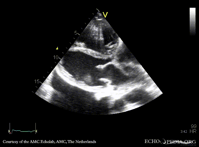
|descriptionfile1=PLAX: prolaps of PMVL, huge amount of pleural effusion | |descriptionfile1=PLAX: prolaps of PMVL, huge amount of pleural effusion | ||
|filepointer2= | |filepointer2=[[File:E00451.gif|350px]] | ||
|file_name2=E00451 | |file_name2=E00451 | ||
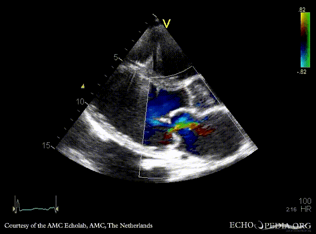
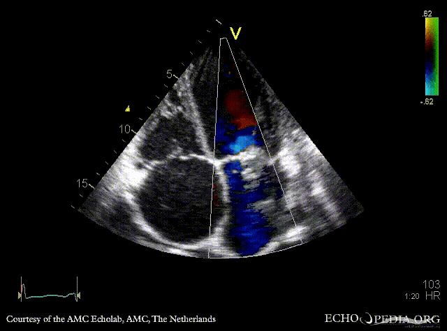
|descriptionfile2=PLAX: Color Doppler, severe mitral regurgitation, excentric jet | |descriptionfile2=PLAX: Color Doppler, severe mitral regurgitation, excentric jet | ||
|filepointer3= | |filepointer3=[[File:E00452.gif|350px]] | ||
|file_name3=E00452 | |file_name3=E00452 | ||
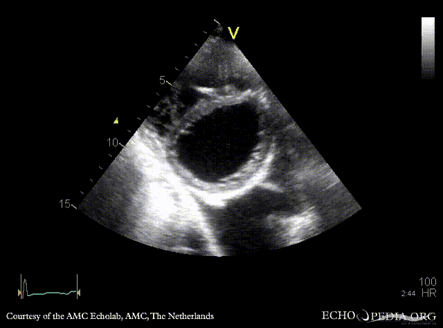
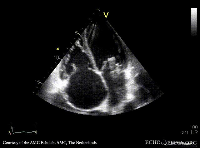
|descriptionfile3=PSAX: flattening of IAS, enlarged right ventricle | |descriptionfile3=PSAX: flattening of IAS, enlarged right ventricle | ||
|filepointer4= | |filepointer4=[[File:E00453.gif|350px]] | ||
|file_name4=E00453 | |file_name4=E00453 | ||
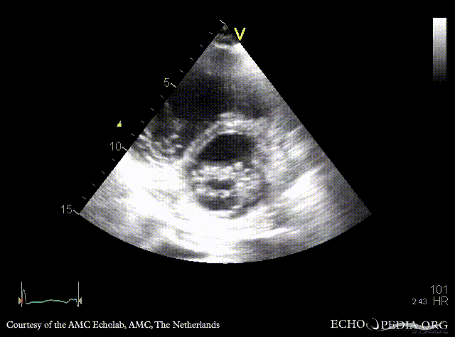
|descriptionfile4=PSAX: prolaps of PMVL | |descriptionfile4=PSAX: prolaps of PMVL | ||
|filepointer5= | |filepointer5=[[File:E00454.gif|350px]] | ||
|file_name5=E00454 | |file_name5=E00454 | ||
|descriptionfile5=A4CH: prolaps of PMVL, enlarged left and right atrium, poor function of dilated right ventricle | |descriptionfile5=A4CH: prolaps of PMVL, enlarged left and right atrium, poor function of dilated right ventricle | ||
|filepointer6= | |filepointer6=[[File:E00455.gif|350px]] | ||
|file_name6=E00455 | |file_name6=E00455 | ||
|descriptionfile6=A4CH: Color doppler, severe mitral regurgitation | |descriptionfile6=A4CH: Color doppler, severe mitral regurgitation | ||
|filepointer7= | |filepointer7=[[File:E00456.gif|350px]] | ||
|file_name7=E00456 | |file_name7=E00456 | ||
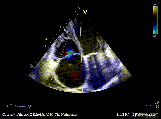
|descriptionfile7=A4CH: moderate tricuspid regurgitation | |descriptionfile7=A4CH: moderate tricuspid regurgitation | ||
Latest revision as of 20:35, 1 December 2023
| Courtesy of: AMC Echolab, AMC, The Netherlands | |

|

|
| PLAX: prolaps of PMVL, huge amount of pleural effusion | PLAX: Color Doppler, severe mitral regurgitation, excentric jet |

|

|
| PSAX: flattening of IAS, enlarged right ventricle | PSAX: prolaps of PMVL |

|

|
| A4CH: prolaps of PMVL, enlarged left and right atrium, poor function of dilated right ventricle | A4CH: Color doppler, severe mitral regurgitation |

|
 |
| A4CH: moderate tricuspid regurgitation | Coninuous-wave signal of moderate tricuspid regurgitation, increased systolic pulmonary artery pressure |
 |
|
| Pulsed-wave signal of pulmonary veins, decreased systolic flow | |