Mitral Stenosis
Quantification of mitral valve stenosis1
| Slight | Moderate | Severe | |
|---|---|---|---|
| PGmean | <5mmHg | 5-10mmHg | >10mmHg |
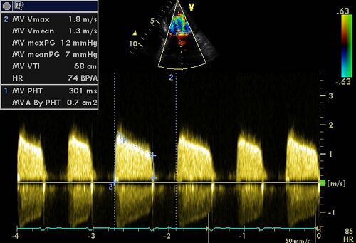
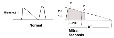
| PHT | <90m/s | 90-150m/s | >150m/s |
| MVA | >1.5cm2 | 1.5-1cm2 | <1cm2 |
Click here to calculate MVA by PHT

|
| Moderate / severe MS |
|---|
Various techniques can be used to estimate the severity of a mitral valve stenosis.
The peak pressure is unreliable for assessing the severity of mitral valve stenosis. Also, the pressure differential is strongly influenced by the heart rate. A high heart rate will greatly increase the pressure difference due to the strongly reduced duration of diastole. For heart rates between 60 - 90/min , the average gradient is estimated by "tracing" of the CW signal across the mitral.
Pressure half-time
Pitfalls:
In any situation where the LV diastolic pressure rises rapidly (AOI, non-compliant LV, myocardial ischemia) it will give an underestimation of the severity of mitral valve stenosis. Immediately after the balloon dilatation of the mitral valve the PHT method is unreliable. In tachycardia at which the A-top merges with the E-top or the atrial flutter waves the E-wave deformation may overestimate the incidence of mitral stenosis.
Heart rates of between 60 and 90/min give best results.
Continuity equation
The continuity equation can be used as the flow through the mitral valve is the same as the flow through the aortic valve and pulmonary valve (mitral valvular insufficiency and aortic valve insufficiency or absence of pulmonary valve insufficiency) .
MVA = TVI(lvot) x area (lvot) / TVI(mv)
Tracing the mitral valve surface
Also when tracing of the mitral valve surface is done, the result of this determination is highly dependent on the gain setting of the ultrasound device, and if it was the smallest opening of the mitral valve is traced. The results, therefore, they are less reliable and reproducible when compared with the calculated mitral valve surface. 3D - echo is more suitable to still make a reliable route, but it is, however, strongly dependent on the picture quality.
Assess suitability mitral plastic
Echocardiography can be predicted whether a patient develops a significant mitral valvular insufficiency after balloon angioplasty. Wilkins was the first to develop a score that the risk of an unsuccessful balloon angioplasty can be estimated.2
Click here for Wilkins score (also called Mitral Valvuloplasty score)
References
<biblio>
- 1 pmid=20435783
- 2 pmid=3190958
</biblio>
